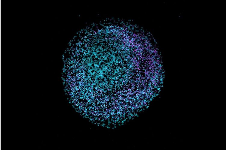
Exo-spin™ blood


Product description
Exo-spin™ products provide a proven, rapid and reliable way of generating high quality purified exosomes suitable for a range of research applications.
Blood plasma and sera typically contain around 1 x 10¹² exosomes/ml. Size exclusion chromatography (SEC), provides a superior to technique to purify exosomes from blood. Blood can be added directly onto the column or concentrated a little, if necessary, prior to application. Blood plasma has higher protein content than sera reducing the amount that can be loaded onto each column.
The EX02 Exo-spin™ kit has been developed to process small volumes of blood samples (both plasma and sera). The kit includes Exo-spin™ buffer as well as SEC columns. The Exo-spin™ buffer is a polymer-based precipitation buffer which can be used for the optional first step. For the second step, pre-packed and equilibrated ready-to-use SEC columns provide reliable and consistent results.
Products on this page contain Exo-spin™ mini columns
Which Exo-spin™ kit do I use for my sample?
Plasma and Sera |
|||
Sample Volume |
isolation method |
Exo-spin Kit |
column bed length |
< 100 µl Note 1 |
SEC |
EX03 mini columns |
1.3 cm |
< 150 µl |
SEC |
EX05 mini-HD | 6.35 cm |
100 µl - 250 µl Note 2 | concentration + SEC | EX02 mini blood | 1.3 cm |
1 ml | SEC | EX04 midi | 2.15 cm |
Note 1: Sera only. Note 2: Up to 500 ul for sera.
Exo-spin™ column and resin pore size
The column bed volume in EX02 Exo-spin™ mini-columns is 500 µl, allowing for 100 µl of volume to be loaded onto each column.
The pore size within the resin is approximatively 30 nm to attain a highly pure exosome elution. All other molecules (e.g. proteins, lipids) which are smaller than 30 nm will enter into the pores and remain trapped in the column.
Highest recovery and purity
Exosomes in a range 30-250 nm will elute in the first fraction. All other proteins and lipids will be retained in the column and will elute later than the exosomes, ensuring a highly pure sample ready for downstream application.
Reproducibility
All our columns are manufactured in our laboratory to ensure a high reproducibility between each lot. As proof of reproducibility and batch-to-batch consistency, a large number of peer-reviewed scientific papers have been published describing the use of Exo-spin™.
Storage
Upon receipt, store purification columns and Exo-spin™ Buffer at 4°C. All other components should be stored at room temperature (15°C - 25°C).
Frequently Asked Questions (FAQs)
For any additional questions, please refer to FAQs document below.
If you are just starting to work with exosomes, consider our starter pack
We designed the ideal starter pack to guide your exosome research. The starter pack includes the exosome purification kit of your choice, exosome validated antibodies, and NTA profiling analysis. Complete details can be found in the product page here.
Product data
EX02 Exo-spin™ kit comparative data
Data determined by nanoparticle tracking analysis (NTA). Each curve represents the average of 3 technical replicate measurements for each exosome isolation method and biofluid triplicate experiment. (PM = Precipitation Method). Figure taken and adapted from (Martins, TS et al., 2018).
Data determined by NTA and EXOCET. Serum sample has been analysed for qualitative comparison between Exo-spin™ and precipitation methods.
Characterizing Exo-spin™ isolated exosomes using nanoparticle tracking analysis (NTA)
Exosome characterization with NTA. Exosomes have been isolated using the Exo-spin™ kit and analysis performed with the ZetaView® instrument.
Downstream applications: advice and content
The EX02 Exo-spin™ kit is compatible with all downstream application and has been published in a large range of different applications.
RNA analysis:
- Wang L., Wang C., Jia X. and Yu J. (2018). Circulating Exosomal miR-17 Inhibits the Induction of Regulatory T Cells via Suppressing TGFBR II Expression in Rheumatoid Arthritis. Cellular Physiology and Biochemistry; 50:1754–1763.
- Emanueli C., Shearn AI., Laftah A., Fiorentino F., Reeves BC., Beltrami C., Mumford A., Clayton A., Gurney M., Shantikumar S. and Angelini G. (2016). Coronary Artery-Bypass-Graft Surgery Increases the Plasma Concentration of Exosomes Carrying a Cargo of Cardiac MicroRNAs: An Example of Exosome Trafficking Out of the Human Heart with Potential for Cardiac Biomarker Discovery. PLoS One 29;11(4):e0154274.
Mass Spectrometry:
- Menezes-Neto A., Fidalgo Sáez MJ., Lozano-Ramos I., Segui-Barber J., Martin-Jaular L., Estanyol Ullate JM., Fernandez-Becerra C., Borrás FE., and del Portillo HA. (2015). Size-exclusion chromatography as a stand-alone methodology identifies novel markers in mass spectrometry analyses of plasma-derived vesicles from healthy individuals. Journal of Extracellular Vesicles; 4: 10.3402/jev.v4.27378.
Functional study:
- Sheller-Miller S, Trivedi J, Yellon SM, and Menon R. (2019). Exosomes Cause Preterm Birth in Mice: Evidence for Paracrine Signaling in Pregnancy. Scientific Reports 24;9(1):608.
Citations
Sheller-Miller S, Trivedi J, Yellon SM, and Menon R. (2019). Exosomes Cause Preterm Birth in Mice: Evidence for Paracrine Signaling in Pregnancy. Scientific Reports 24;9(1):608.
Soares Martins, T., Catita, J., Martins Rosa, I., Cruz da Silva, O., and Henriques, A.G. (2018) Exosome isolation from distinct biofluids using precipitation and column-based approaches. PLoS One. 13(6):e0198820.
Wang L., Wang C., Jia X. and Yu J. (2018). Circulating Exosomal miR-17 Inhibits the Induction of Regulatory T Cells via Suppressing TGFBR II Expression in Rheumatoid Arthritis. Cellular Physiology and Biochemistry; 50:1754–1763.
Emanueli C., Shearn AI., Laftah A., Fiorentino F., Reeves BC., Beltrami C., Mumford A., Clayton A., Gurney M., Shantikumar S. and Angelini G. (2016). Coronary Artery-Bypass-Graft Surgery Increases the Plasma Concentration of Exosomes Carrying a Cargo of Cardiac MicroRNAs: An Example of Exosome Trafficking Out of the Human Heart with Potential for Cardiac Biomarker Discovery. PLoS One 29;11(4):e0154274
Menezes-Neto A., Fidalgo Sáez MJ., Lozano-Ramos I., Segui-Barber J., Martin-Jaular L., Estanyol Ullate JM., Fernandez-Becerra C., Borrás FE., and del Portillo HA. (2015). Size-exclusion chromatography as a stand-alone methodology identifies novel markers in mass spectrometry analyses of plasma-derived vesicles from healthy individuals. Journal of Extracellular Vesicles; 4: 10.3402/jev.v4.27378.