EV MISEV Service Package
Service Package outline
- Isolation of EV/exosome content using Exo-Spin
- Measurement of total protein content using a BCA assay
- Measurement of EV/exosome numbers and size distribution using nano tracking analysis (ZetaView)
- Lipid quantification
- Western blot against three tetraspanin markers
- Biomarker quantitative analysis: ExoLISA assay with three tetraspanin markers
- Scanning electron miroscopy (optional extra)
Service Requirements
Samples shipped either as live cell cultures or dry ice shipments.
- Cell Culture min 10 ml max 500 ml
- Plasma/sera - Min 0.1 ml. Max 1 ml
Please supply details of your samples using the exosomes service enquiry form
| Product Details | |
|---|---|
| Service Duration |
1 - 2 weeks from receipt of samples |
| Yields |
Sample dependent. Typically: cell culture media (normal cells) 109 exosomes per ml |
Service Components
This is a comprehensive service for the purification and proteomic analysis of exosomes.
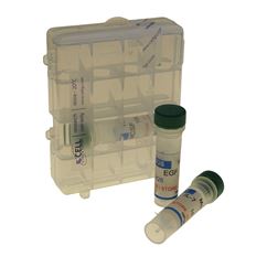
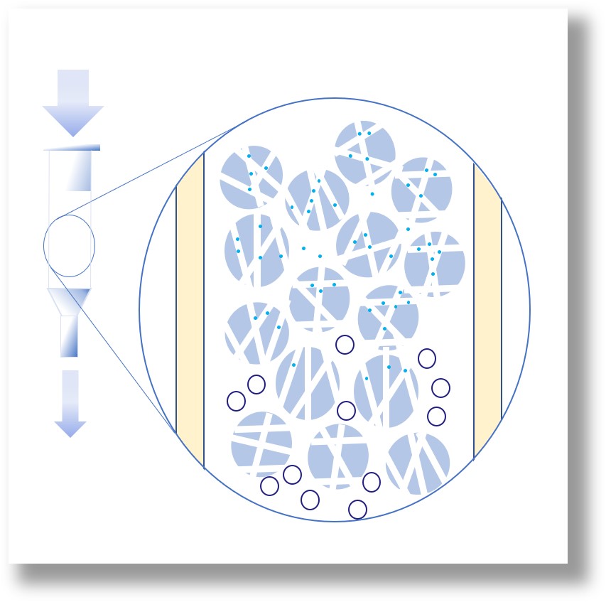
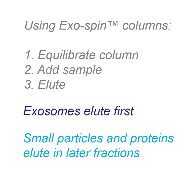
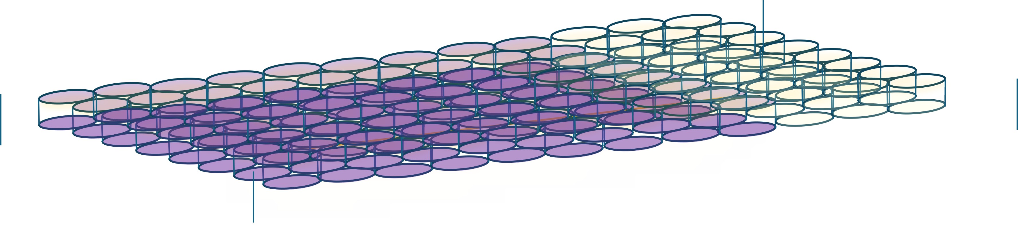
1. Isolation of EV/exosome content using Exo-Spin size exclusion chromatography (SEC)

The sample is spun to remove cells and (optional step) precipitated to concentrate exosomes. (2) The column closures are removed (3) the column is equilibrated (4) the sample is added (5) purified exosomes are eluted
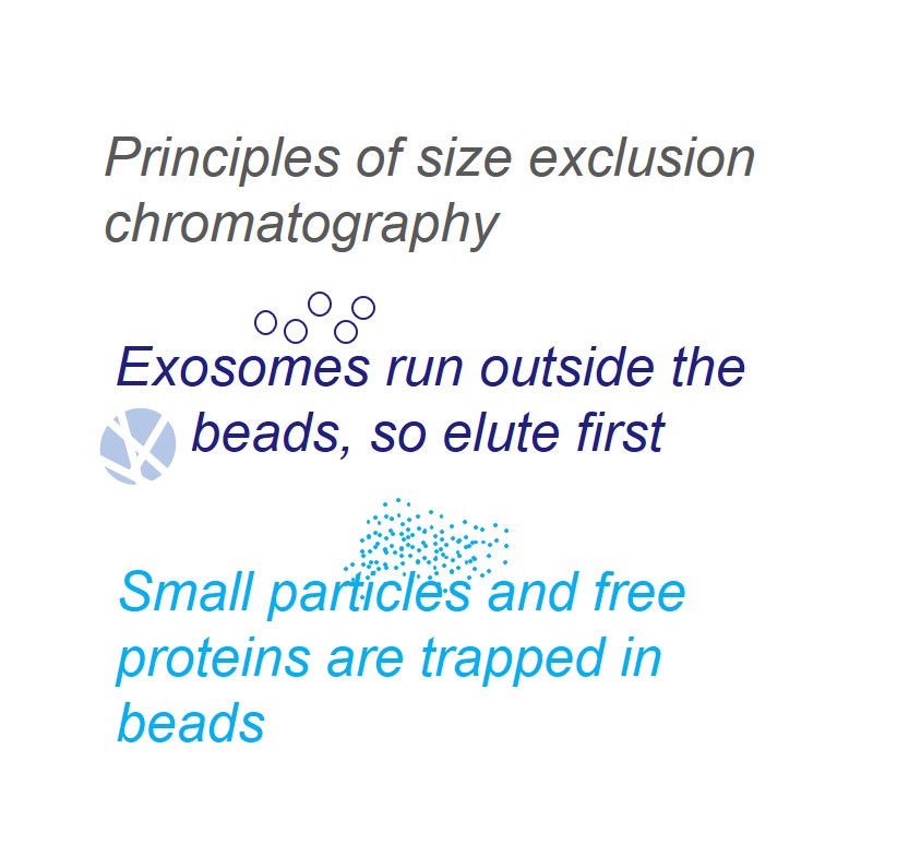
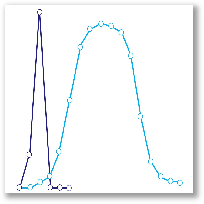
SEC separates particles in solution based on their size. An SEC column is packed with resin containing stable polymeric spherical beads, each of which has large numbers of nano-scale pores across its surface. The beads leave gaps after packing, the size of which depends on the diameter of the beads. When a solution containing exosomes is added to the top of the resin bed, the smallest particles, including free-proteins, become trapped in the nano-sized pores, slowing their progress, whereas the larger exosomes pass around the outside of the beads and elute first. The very largest particles, beyond the size of the gaps between the beads, are also slowed or stopped completely. The resin used in Exo-spin columns has been chosen to specifically allow primary elution of particles in the range 30nm - 250nm. This allows the isolation of exosomes from particles of other sizes.
2. Measurement of total protein content using a BCA assay
- Copper (II) is reduced to copper (I) in an alkaline solution
- Bicinchoninic acid binds to the copper (I)
- The BCA-copper complex turns purple
- The amount of purple color is proportional to the amount of protein in the sample
Colorimetric assay for protein content
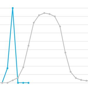
3. Measurement of EV/exosome numbers and size distribution using nano tracking analysis (ZetaView)
Cell Guidance Systems service uses ZetaView®, a second-generation instrument from Particle Metrix GmbH, which performs rapid in vitro measurement of physical parameters, including size, surface charge, and concentration of particles.
The ZetaView® instrument has been designed for automated scanning NTA. To provide a more representative analysis, the software measures 11 distinct positions in each sample. This provides more detailed information than single-point assessments, facilitating repeatable and reproducible results. Furthermore, automated parameters for auto-alignment and autofocus ensure that the video quality is kept constant as the particle movement is recorded.
Zetaview NTA technology is well suited for exosome characterization, as it allows for easy and quick measurements, and is compatible with low-concentrated and polydisperse samples.
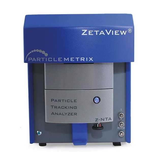
ZetaView® instrument used for the NTA exosome profiling service
4. Lipid Quantification
We use a robust assay to analyze the lipid content of your sample. The lipids in EV samples primarily come from the EVs themselves. This assay provides supportive evidence for EV numbers and can be used to augment NTA analysis.

Colorimetric assay for lipid content
5. Western blot
For human samples only. We use validated antibodies to check for common tetraspanin markers CD9, CD63 and CD81
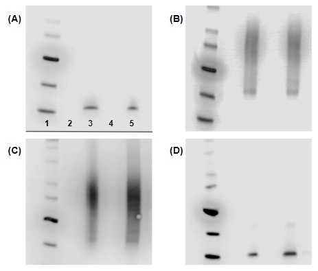
Western blot images generated using cell lysate from DU145 cells (human prostate cancer cell line). The cell lysate was loaded preceding the exosome sample detected with (A) CD9 mAb (clone CGS12A mAb), (B) CD63 mAb (clone CGS73B), (C) CD63 mAb (clone CGS82X), and (D) clone CGS36K mAb. The content of each lane is as follows: Lane 1 – Invitrogen™ Magic Mark™ ladder; lane 2 – 1 µg cell lysate; lane 3 – 1 µg exosome protein; lane 4 – 2 µg cell lysate; lane 5 – 2 µg exosome protein. The mAb concentration used to generate this western blot was 0.5 µg/ml or a 1:2000 dilution of the 1 mg/ml stock solution.
6. Exosome Biomarker analysis
In addition to Western blotting, the ELISA-like ExoLISA assay can detect markers with exquisite sensitivity using Europium time-resolved fluorescence
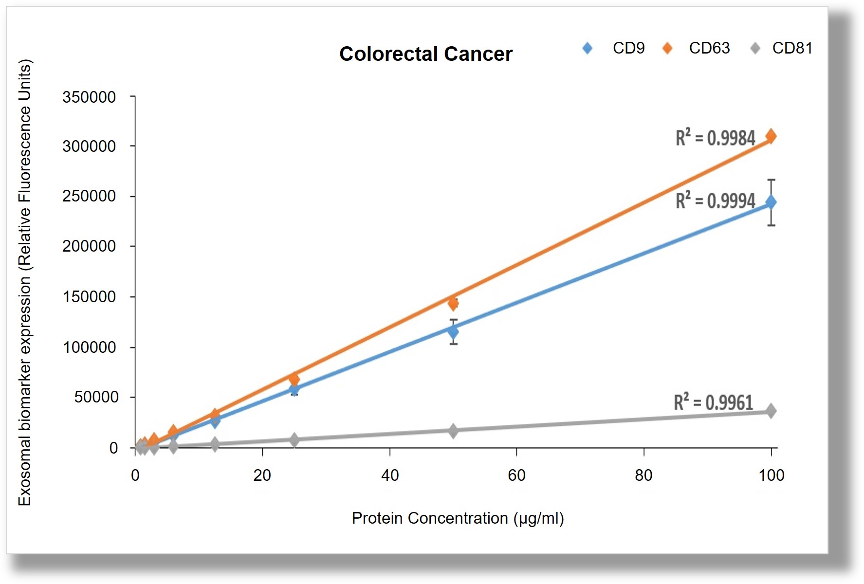
Differential expression of exosomal markers from cultured colorectal cancer cells detected in an ExoLISA assay. For further details please see the product page
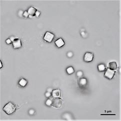
7. Transmission electron miroscopy (optional extra)
Purified exosomes imaged on a Zeiss 550 Crossbeam microscope.
This analysis is the best MISEV practice:
- Confirms the characteristic size and cup-shaped or round morphology of exosomes
- Distinguishes EVs from protein aggregates or other particulates
- Verifies structural integrity of the vesicles


Zeiss 550 Crossbeam SEM imaging of exosomes.
Ask for a quote
Please get in touch if you have any questions via the chat box on the right of this page or email [email protected]