PeptiGel® Technology
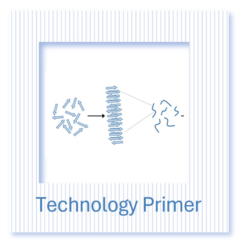


Next-generation self-assembling peptide hydrogel (SAPHs)

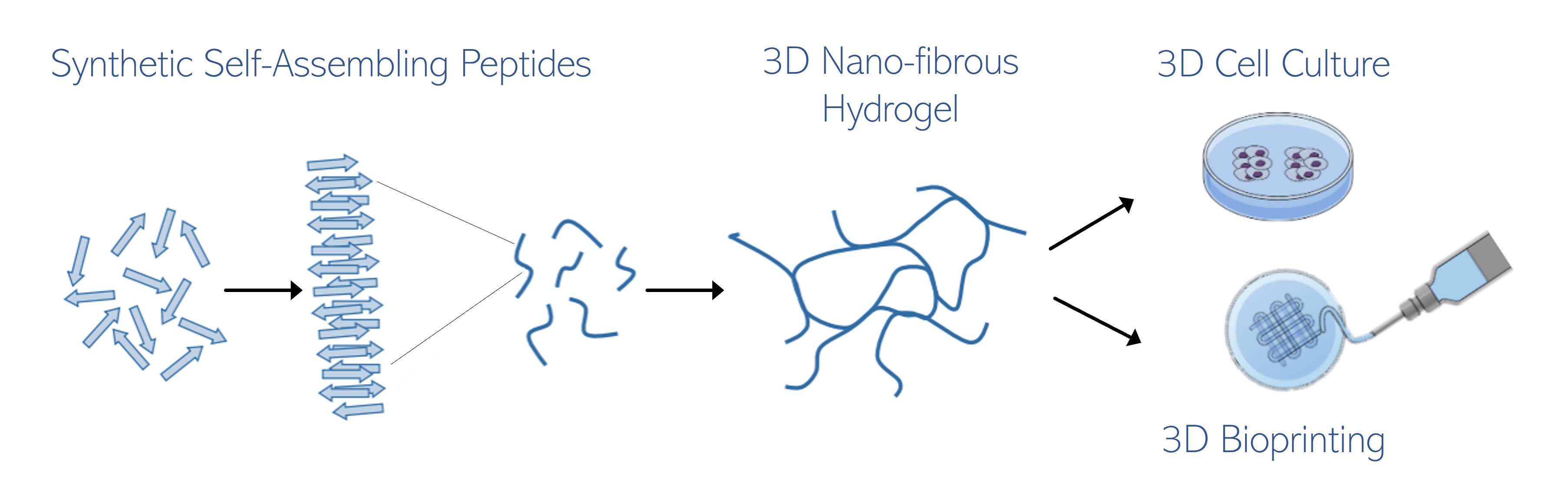
Short synthetic peptides self-assemble into stacks that develop into 3D nano-fibrous matrix hydrogel networks
Meet the Family
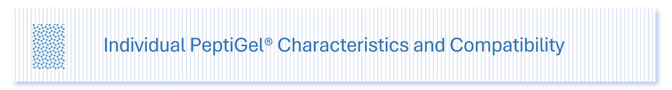
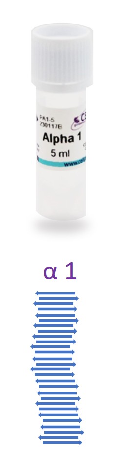
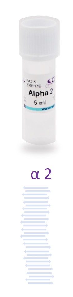
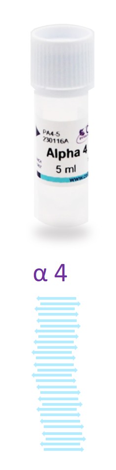
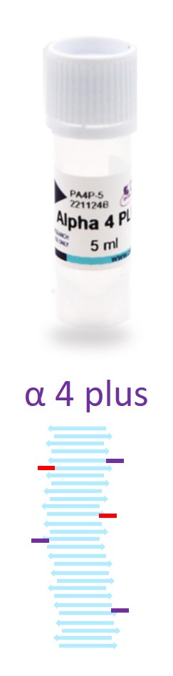

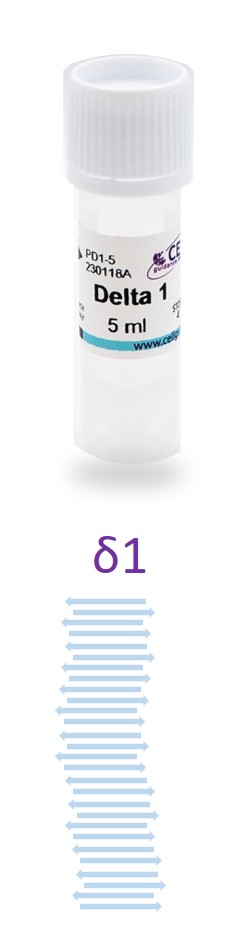
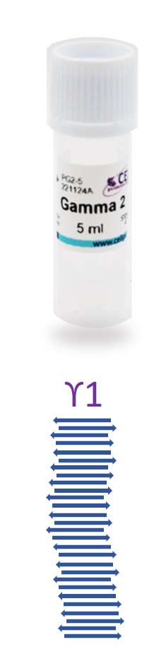
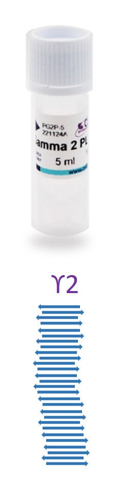
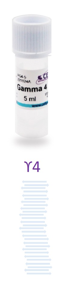
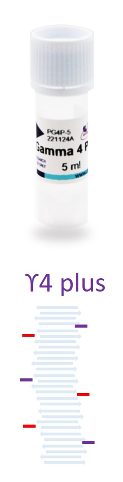
The PeptiGel family. Finely tuned (for charge, elasticity) peptides, which may be functionalized (PLUS versions) with bioactive peptide motifs
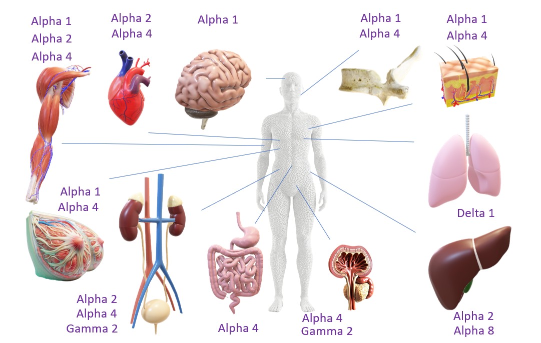
Tailored for each tissue and cell type
PeptiGel is a family of related hydrogel formulas that provide a range of class-leading SAPHs and bioinks to support your cell culture applications. The proprietary hydrogel technology allows us to tune mechanical and functional properties, to provide you with the most suitable materials tailored to your application's needs.
- The peptide backbone sequence is modified affecting properties such as charge and elasticity.
- Bioactive peptide motifs, such as RGD and GFOGER are fused to the main sequence, completely integrating them into the hydrogel.
Of course, PeptiGels may be diluted to optimize viscosity. You can be assured that PeptiGels® are physiological and biologically relevant hydrogels that mimic the cell micro-environment and provide a synthetic extracellular matrix.
Click the image above to see the growing range of successfully cultured cell types.
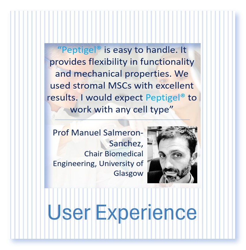
PeptiGel® — What Researchers Say
Many types of cells and tissues have been cultured with PeptiGel®
Validated Across 40+ Cell Types
PeptiGel® synthetic peptide hydrogels have been independently validated across more than 40 cell types, spanning stem cells, primary cells, immortalised cell lines, and patient-derived material. Validated applications include:
- Stem cells: mesenchymal stem cells (MSCs), induced pluripotent stem cells (hiPSCs), neural stem cells, adipose-derived stem cells (dDASCs)
- Cancer models: pancreatic ductal adenocarcinoma (PDAC), MCF-7 breast cancer cells, epithelial ovarian cancer cells, primary tumour cells
- Organoids: kidney organoids (hiPSC-derived), liver organoids, gastrointestinal organoids
- Connective tissue and musculoskeletal: synoviocytes, chondrocytes, osteoblasts, stromal fibroblasts (rOSFs)
- Cardiovascular and vascular: cardiomyocytes, human umbilical vein endothelial cells (HUVECs)
- Neural: Schwann cells, Schwann cell-like dDASCs, neural progenitor cells
- Epithelial: mammary epithelial cells, kidney tubular epithelial cells
Each PeptiGel® variant can be matched to your target cell type by tuning stiffness, charge, and bioactive functionalisation (RGD, IKVAV, YIGSR, GFOGER). View the full validated cell line table, or request a bespoke PeptiGel® design for your specific application.
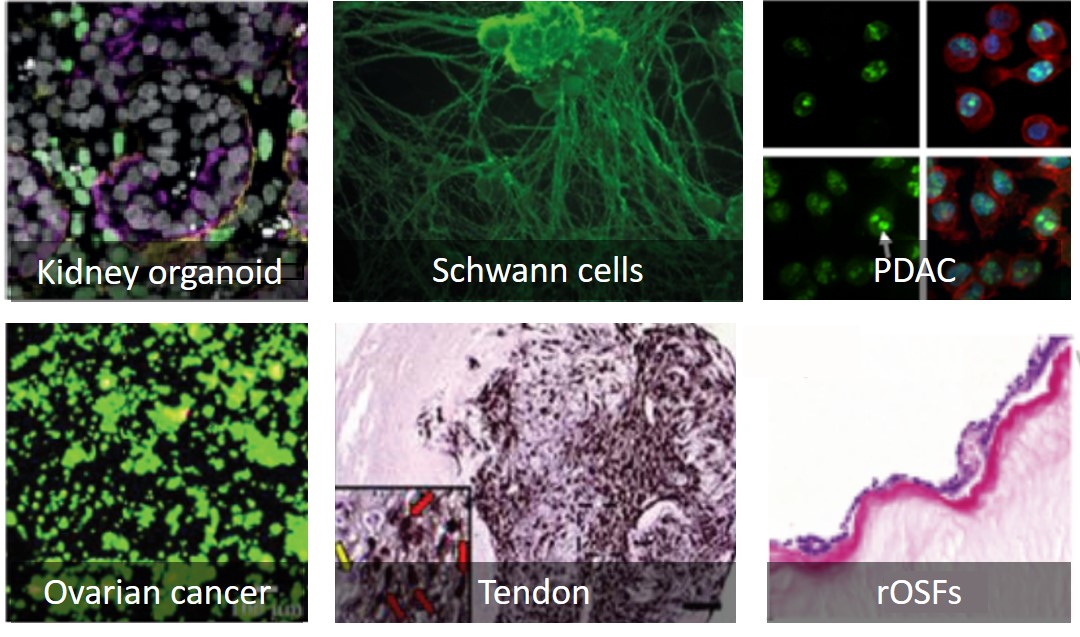
Examples of cells grown with PeptiGel® hydrogels. PDAC= pancreatic ductal adenocarcinoma, rOSFs = rat oesophageal stromal fibroblasts.
Bespoke Peptide Hydrogel Design
The chemistry, mechanical and bio-functional properties of each PeptiGel® can be tuned to create bespoke products to suit your cells’ needs. This includes functionalizing a PeptiGel® with peptide sequences from collagen (GFOGER), fibronectin (RGD), & laminin (IKVAV or YIGSR). We can also tailor the material properties to precisely fit your requirements for a wide range of application areas, including cell therapies, drug delivery, and high-throughput screening.
If you require specific material design characteristics or assistance choosing the PeptiGel® that's most suitable for your application, please get in touch.
Key Technology Benefits
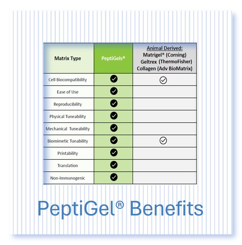
PeptiGels® peptide hydrogels are fully synthetic and spontaneously self-assemble to form 3D nano-fibrous hydrogels that mimic the native extracellular matrix (ECM). The mechanical stiffness of these hydrogels is modulated and matches the stiffness of most tissue types. The fibre surfaces can be (bio)chemically functionalised with several biomimetic peptide sequences from key ECM proteins that are proven to signal and enhance biological processes. These include RGD (fibronectin), IKVAV (laminin), YIGSR (laminin) and GFOGER (collagen). The ability to tune the properties of the peptide hydrogels to provide the optimal environment for your cells’ needs, makes them the ideal synthetic alternative to animal-derived matrices such as Matrigel™, Geltrex™ and collagen. As peptides are the building blocks of nature, PeptiGels® are inherently biocompatible and provide a suitable environment for cells to survive and thrive
- Mechanically tuneable - PeptiGels® come in a range of mechanical strengths and viscosities,s enabling you to optimise the scaffold for your cells’ and bioprinting needs. The range of stiffness available mimics all human tissue types and provides control over cell behaviour and fate. PeptiGels are slightly viscous and further solifdify in the presence of cell culture media.
- Biochemically functional - Bioactive peptide sequences (such as RGD and GFOGER) can be fused to the core morif and incorporated within peptide hydrogels to influence the dynamic interplay between cells and the ECM.
- Animal and pathogen free - PeptiGels® are fully synthetic and animal product free. They are 100% ethical. They are formulated to closely mimic a range of human tissues and in vivo environments, thus increasing translatability to humans.
- Resorbable - Since PeptiGels® are peptides, they are degraded by proteases, such as matrix metalloproteinase. The gel is completely adsorbed within a few weeks. This property also allows cell migration.
- Reproducible - PeptiGels® are manufactured in a dedicated facility with rigorous quality control, ensuring consistent product quality. This gives you confidence that you will achieve the same results each time, providing enhanced data quality and reliability.
- Convenient and ready-to-use - PeptiGels® are supplied as ready-made semi-vicous hydrogels. There are no complex temperature or pH control steps required; you simply seed your cells at room temperature and get consistent results, saving you both time and hassle.
- Sprayable, injectable and printable - The shear-thinning properties of our hydrogels enables their flexible handling, opening up their use in high-throughput liquid handling systems and extrusion-based bioprinting. It also enables versatile delivery by injection, spray or endoscope for targeted drug delivery and cell therapies.
- Transparent - PeptiGels® are optically transparent making them compatible with imaging techniques to allow for live-cell imaging and real-time quantification of cell mobility.
- Clinically translatable - PeptiGels® are fully defined and their inherent biocompatibility (they are simple peptides) enables clinically translatable research.
Webinar: How to Successfully Make the Switch to Synthetic Peptide Hydrogels
Professor Aline Miller (University of Manchester) and Sebastian Doherty-Boyd (University of Glasgow) discuss the difficulties in producing consistent cell culture results using traditional biomaterials, particularly animal-derived matrices with their inherent variability. They also discuss how to gain greater control over 2D and 3D cell culture with PeptiGels and cover specific examples of using PeptiGels to generate 3D tissue and disease models. Published: September 2023
Comparison of 2D cell culture and 3D cell culture with PeptiGel®
PeptiGels® can be used for 2D and 3D cell culture, and have been proven successful in supporting a range of application areas. See how they compare.
2D vs. 3D cell culture |
|
2D Cell Culture |
3D Cell Culture |
| Not representative of the in-vivo environment | Better simulation of the in-vivo environment |
| Altered cell-to-cell interactions and signalling | Enhanced cell-to-cell interactions and signalling |
| Need for animal testing for validation | Reduction in animal usage |
| Limited in its application areas | Wide-ranging applications e.g. integration of fluid flow and bioprinting |
| Lack of in-vivo predictivity | Reliable and relevant results |
| Simple to analyse | Improved method to model diseases |
| Well established | Not as widely explored |