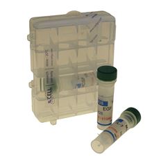
Exosome CD antigen antibody selection
Product description
A collection of validated antibodies against tetraspannin CD antigens CD9, CD63, and CD81.
Reactivity
Each CD antibody is cross-reactive against human and mouse CD proteins
The exosome antibody has been validated against exosome-associated antigens to characterize and/or quantify exosomes in cell culture media and biological fluids, making them an essential tool in exosome research.
All high-quality exosome validated antibodies have been selected to specifically detect CD9, CD63, and/or CD81 which are widely used as exosome markers. The antibodies can be selected individually or as part of a convenient and economical collection containing 20 µg of each of the following antibodies: CD9 Clone CGS12A, CD63 Clone CGS82X, and CD81 Clone CGS36K.
Western Blotting
Western blotting is one of the most common methods to assess the presence of specific proteins in extracellular vesicles (EV) samples. In order to determine if the analyzed proteins are enriched in exosomes, the western blotting is typically loaded with exosome samples side-by-side with source material lysates.
ExoLISA™ detection assay
The exosome marker antibodies are the same as the ones used in the ELISA-like detection assay, please see full details of the ExoLISA™ kit here.
Storage
Upon receipt, store antibodies at 4°C.
Product data
Western blotting


Western blot images generated using A375 (Human melanoma) and mouse serum-derived exosomes isolated with Exo-spin™ MIDI columns (EX04). The CD mAb concentrations used to generate this western blot was a 1:1000 dilution of the 1 mg/ml stock solution.
Human: Lane 1 and 3 – Invitrogen™ MagicMark™ ladder; lane 2 – 20 µg exosomes from Exo-spin™ MIDI fractions 7-12 (pooled).
Mouse: Lane 1 and 3 – Invitrogen™ MagicMark™ ladder; lane 2 – 30 µg exosomes from Exo-spin™ MIDI fractions 7-12 (pooled).
References
Kosanovic M, Milutinovic B, Goc S, Mitic N, Jankovic M. Ion-exchange chromatography purification of extracellular vesicles. (2017). Biotechniques 63(2): 65-71.


