RGD (Arg-Gly-Asp) peptide, 5 mg
Description
RGD peptide is a synthetic peptide containing the RGD cell attachment sequence found in fibronectin, vitronectin and many other matrix and serum proteins.
Why our RGD is better
The binding of this peptide is mediated via a hydroscopic C-terminal sequence fused to the RGD motif, providing a multi-modal mechanism of action which optimizes cell attachment and activation via integrin receptors. This RGD peptide has been specifically engineered to spontaneously adhere to various surfaces such as polystyrene (either tissue culture treated or non-treated), hollow fibres made of cellulose, glass, polycarbonate membranes and many other materials. This enables enhanced use in a wide range of cell culture applications.
Research Use Only
Procedures
Please note: Use these recommendations as guidelines to determine the optimal coating conditions for your cultures system. Two options are provided: procedure A and procedure B.
Procedure A
- Remove cap and add 5 ml of serum-free medium or PBS to the bottle.
- Replace cap and vortex contents vigorously. Ensure that the RGD peptide is completely solubilized. The solution will remain slightly hazy.
- Transfer desired volume of solution from the bottle to a dilution vessel. Dilute to desired concentration using serum-free medium or PBS. A typical working concentration may range from 0.1 to 10 μg/ml.
- Sterile filter solution through a 0.22 micron button filter.
- Aseptically add appropriate amount of diluted, sterile material to culture surface.
- Incubate at room temperature or 37°C, covered, for 1 - 2 hours.
- After incubation, aspirate remaining material.
- Rinse plates carefully with dH2O - avoid scratching bottom surface of plates.
- Plates are ready for use. They may also be stored at 2 - 10°C damp or air dried if sterility is maintained.
- Store remaining solubilized RGD peptide at 2 - 10°C. Additional note: Include divalent cations (Calcium, Magnesium, or Manganese) in cell attachment solution to obtain optimum cell binding.
Procedure B
- Remove cap and add 5 ml of sterile 70% ethanol.
- Replace cap and vortex contents. Ensure that the RGD peptide is completely solubilized.
- Transfer desired volume of solution from the bottle to a dilution vessel. Dilute to the desired concentration using 70% ethanol. Concentrations from 0.1 to 10 μg/ml should be tested.
- Add appropriate amount of diluted material to culture surface.
- Leave the coated container, uncovered, in a laminar flow hood until the wells are dry.
- Rinse plates carefully with dH2O - avoid scratching bottom surface of plates.
- Plates are ready for use.
- Store remaining solubilized RGD peptide at 2 - 10°C.
| Product Details | |
|---|---|
| Source |
Synthetic |
| CAS Number |
99896-85-2 |
| Counter Ion |
Acetate |
| Purity |
>95% |
| Identity confirmed by amino acid analysis |
Characteristic |
| Identity confirmed by mass spectrometry |
Characteristic |
| Peptide content confirmed by nitrogen analysis |
Characteristic |
| Physical Appearance |
Powder |
| Sterility |
Sterile |
| Cell Attachment Assay |
Pass |
| Peptide Sequence |
Ac-Gly-D-Arg-Gly-Asp-Ile-Pro-Ala-Ser-Ser-Lys-Gly-Gly-Gly-Gly-Ser-D-Arg-Leu-Leu-Leu-Leu-Leu-Leu-D-Arg-NH2 |
| Storage and Stability |
Store at 4°C. |
Mechanism
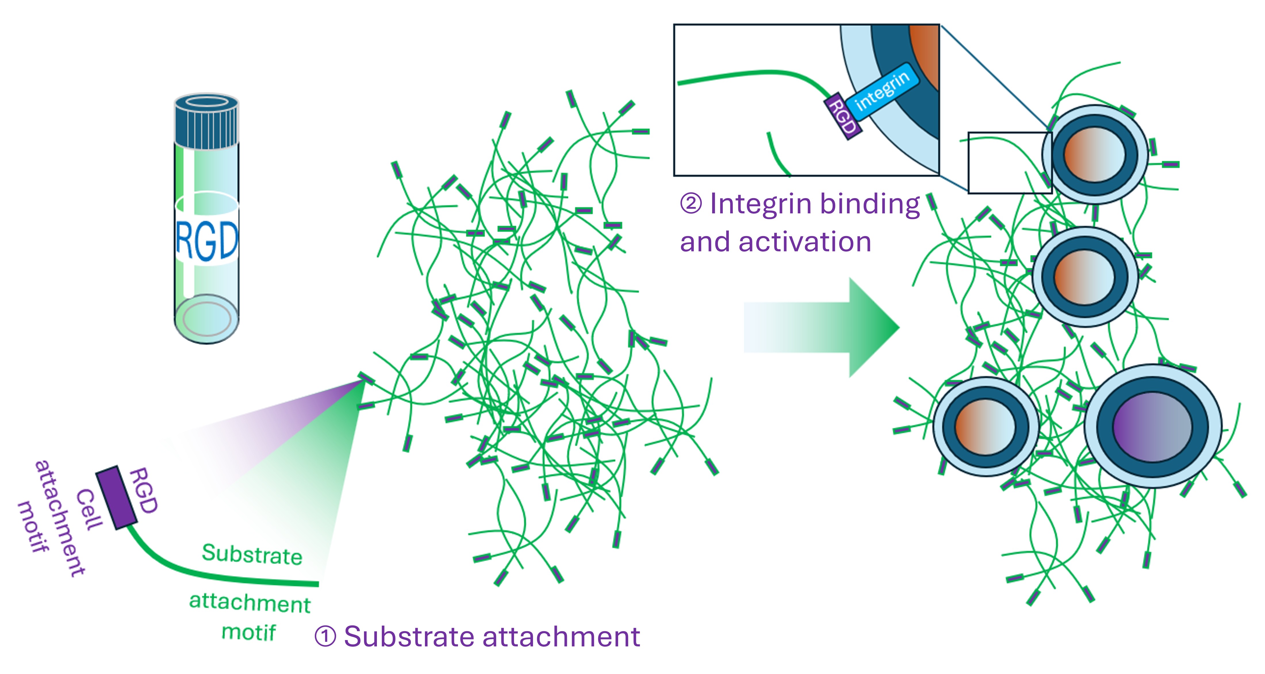
How Substrate affinity tag binds to both the (1) substrate and (2) cells via integrin attachment generating activation
References
Glass, J.R., Craig, W.S., Dickerson, K. et al. Cellular Interaction with Biomaterials Modified by Arg-Gly-Asp Containing Peptides. MRS Online Proceedings Library 252, 331–337 (1991). https://doi.org/10.1557/PROC-252-331