An easier way to model biological gradients

Protein gradients, especially of cytokines and chemokines, are essential in biology for guiding cell migration, activating immunity, and orchestrating development. Traditional methods often struggle with unstable delivery, burst release, or limited spatial control. Here, we review common approaches and highlight the befits of PODS as a powerful, next-generation tool for engineering reliable protein gradients.
Why Cytokine Gradients Matter
Gradients of bioactive proteins, notably chemokines (like CXCL12), cytokines (like IL‑2, IFN‑γ), and other grow factors (like TGF‑β, and VEGF) are critical in mammalian biology as they:
- Drive chemotaxis in immune and stem cells.
- Pattern tissues during morphogenesis.
- Tune stem-cell differentiation and tumor microenvironments.
- Maintain tissue structure (homeostasis)
Achieving controlled and sustained delivery of these factors experimentally in vitro or therapeutically in vivo remains a major technical challenge.
| Feature | Traditional Methods | PODS® Delivery System |
|---|---|---|
| Gradient Stability | Short (hours to days) | Long (weeks; steady-state release) |
| Protein Protection | Limited stability | High: protects from degradation & heat |
| Spatial Control | 2D-focused, limited in 3D | Precise 2D/3D placement via printing/scaffolds |
| Integration with Biomaterials | Requires optimization | Built-in microsystem for embedding |
| Example Applications | Simple migration assays | Vascular grafts, organoids, osteogenic grafts |
Traditional Gradient Methods: Pros & Cons
- Microfluidic Devices – offer precise, dynamic gradients, but are often complex and challenging to scale for routine lab use.
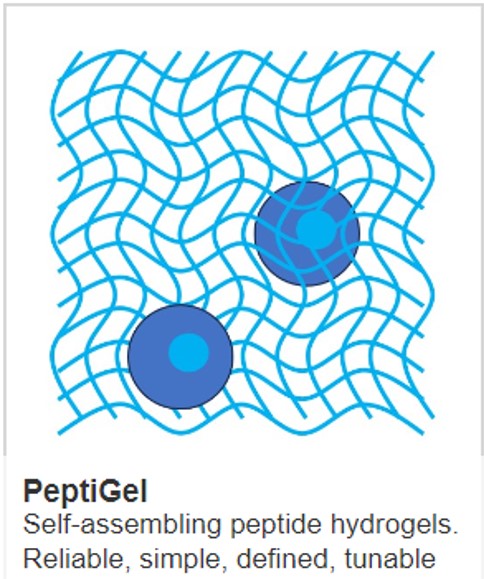
- Hydrogel Embedding – 3D protein-loaded gels support tissue-like culture but suffer from burst release and short gradient lifetime.
- Transwell Diffusion Systems – simple to set up, yet diffusion rapidly dissipates any stable gradient.
- Immobilization/Surface Patterning – yields stable 2D gradients but lacks 3Dity and dynamic modulation.
PODS®: A Smart Delivery Platform
PODS® are micron-sized crystalline protein depots formed by co-expressing polyhedrin with cargos (e.g. cytokines) in insect cells. These crystals slowly degrade via proteases, steadily releasing active proteins over days or weeks while protecting them from degradation.
Key Features of PODS®
- Steady, prolonged release: near zero-order kinetics over several weeks.
- Spatial placement: crystals can be precisely positioned via pipetting or 3D printing, enabling localized gradients.
- Stability & protection: cargos remain biologically active and protected from thermal or enzymatic degradation.
- Biomaterial integration: compatible with hydrogels, scaffolds, or microcarriers for versatile tissue applications.
- Cytokine toolbox: ready-to-use PODS® exist for IL‑3, BMP-2, CXCL12, IL‑6, IFN-γ, TNF-α and more.
Evidence Supporting PODS®
- Phagocytosed Polyhedrin–Cytokine Cocrystal Nanoparticles by Cell and Gene Therapy Insight details nanoscale PODS® loaded with cytokines, showing sustained delivery and proof-of-concept functionality.
- Accelerating vascular graft development demonstrates spatial gradient creation by placing PODS near or far from stem cells in grafts, affecting differentiation outcomes.
- Optimizing Delivery of Therapeutic Growth Factors cites PODS® (e.g. BMP-2) as a robust carrier that enhances bone regeneration via sustained release.
- Sustained Neurotrophin Release explains how polyhedrin crystals (PODS) reliably support neurotrophin delivery, underscoring sustained release in neural tissue models.
Applications & Experimental Use-Cases
- Chemokine Gradients: Embed CXCL12-loaded PODS® in 3D collagen gels to drive directional migration of immune cells toward defined zones.
- Stem Cell Differentiation: Localized TGF‑β, BMP‑2, or BMP‑7 gradients guide MSCs toward osteogenic or chondrogenic lineages, as evidenced in bone repair models .
- Neurobiology & Organoids: Placement of PODS® loaded with BDNF/GDNF supports neurogenesis in retinal organoid cultures with fewer media changes
Practical Tips for Gradient Experiments
- Titrate crystal density to shape release kinetics and gradient range.
- Strategic placement within gels or scaffolds allows directional gradient formation.
- Quantify release using ELISA or reporter assays to confirm spatial concentration.
- Pair with live cells—monitor migration, differentiation, or signalling over time.
IMAGE Protein gradient formation with PODS CREDIT CellGS
Learn more about powerful technologies that are enabling research: