Extracellular Matrix for the Study of Breast Cancer

The extracellular matrix (ECM) is one of the major structural components of the tumour microenvironment, as it is made up of a network of biochemically different components such as fibrous proteins, glycoproteins, proteoglycans and polysaccharides. This makes its structure highly dynamic with various ECM components being deposited, modified or degraded on a regular basis, and the structure undergoing constant remodelling.
The fibrous proteins within the ECM tend to be collagens, which have a huge role to play in tissue development as they alter cell adhesion, promote cell migration and provide mechanical strength. The glycoproteins help to make the ECM a more cohesive network of molecules as they encourage cells to link together through the use of structural components such as fibrillin, fibronectin, fibulin and laminin. Proteoglycans are glycosylated proteins that are present in the ECM of connective tissues – they are made up of a core protein and one or more covalently attached sulfated glycosaminoglycan chains. And finally, polysaccharides buffer physical stress on the ECM and fill in the interstitial space, through the use of a chain of monosaccharide repeats linked through glycosidic bonds.
ECM and the in vivo environment
In the past, the majority of cell-based assays have been based on traditional two-dimensional (2D) monolayer cells that are cultured on flat and rigid substrates. Although this has proven to be a valuable method for many cell-based studies, this also has some inherent limitations with its accuracy in modelling disease. Almost all cells in the in vivo environment, for example, are surrounded by other cells and the extracellular matrix in a three-dimensional (3D) way – and 2D cell culture is not able to take into account this natural 3D environment of cells. This means that quite often, 2D cell culture tests can provide information that is misleading and non-predictive for many in vivo responses.
3D cell culture systems that use the ECM, are therefore more representative of the actual microenvironment where breast cancer cells reside in human tissue, and so, therefore, more accurately represent in vivo cellular responses.
The importance of scaffold stiffness in cell behaviour
Stiffness is crucial when it comes to choosing the most appropriate scaffold, as it has a direct effect on cancer progression and drug resistance. This means it is vital to match the scaffold stiffness as closely as possible to the tissue type in order to ensure the in vivo environment is as representative as possible.
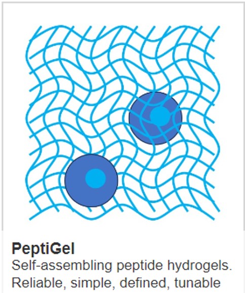
There are a lot of ECM products on the market for researchers to choose from, but what makes PeptiGels, a related family of self-assembling polypeptide hydrogels (SAPHs) so unique is that they are designed with a range of mechanical properties and chemical functionalities to closely mimic the native cellular microenvironment – allowing the growth of any cell type. Their charge, functionality and stiffness can all be independently controlled to replicate the native cellular environment of most human tissue, such as breast tissue, more closely.
In order to show how our innovative peptide hydrogels can support the study of breast cancer, researchers at the University of Manchester published the paper: “Neutrally Charged Self-assembling Peptide Hydrogel Recapitulates in vitro Mechanisms of Breast Cancer Progression” which demonstrated that PeptiGel Alpha1 SAPH can support the growth and expansion of two epithelial breast cancer cell lines, representing different stages of tumours. The researchers also found that Alpha 1 is more suited to studying the early stages of breast cancer than it is for studying metastatic breast cancer.
IMAGE - PeptiGel
Learn more about powerful technologies that are enabling research: