How 3D bioprinters are advancing cancer research

Cancer is the second leading cause of death worldwide, after cardiovascular disease, with over 16.4 million cancer-related deaths predicted by 2040. It is well known for its complex and dynamic nature, which has meant that it is challenging to study with 2D cell culture models, although understanding of the disease has improved.
However, the advent of 3D cell culture, specifically 3D bioprinting, has positively impacted cancer research. It has meant that the more complex aspects of cancer can be modelled and studied, such as metastasis. Researchers can also test cancer-related drugs’ efficacy and toxicity in a more realistic environment.
Research by Vanderburgh, Sterling and Guelcher published in the Annals of Biomedical Engineering in 2016 revealed that cells grown in 3D mimic in vitro cellular responses more accurately.
How does 3D bioprinting work?
3D bioprinters work in the same way as regular 3D printers, except that they deposit layers of biomaterial such as living cells rather than materials such as metal or plastic. The biomaterials, such as kidney or skin cells, are harvested from a living patient and then cultivated to create the bioink.
3D bioprinting also relies on organic or synthetic glue cells that can attach to and grow, such as a collagen scaffold or dissolvable gel. This helps them to mould into the correct form and keeps them stable. The cells then seek out similar cells to join using their inherent properties. Researchers can then control their desired shape, and the printer builds the final structure.
3D bioprinting and cancer research
As 3D bioprinting allows for the layer-by-layer depositing of bioinks, it brings many benefits to cancer research, including:
- Fine-tuning of matrix properties – Many extracellular matrices (ECM) mechanics, such as scaffold stiffness, also play a role in cancer cells’ metastatic behaviour. So, one of the benefits of bioprinting is that this factor can be easily incorporated into a 3D bioprinted tumour model.
- Integration of perfusable vascular networks – A scaffold-based approach to bioprinting allows perfusable vascular networks to be incorporated into the 3D scaffold so cancer cells’ invasion through vessels can be studied.
- High-throughput screening of cancer models – Automation and high-throughput assay capabilities are two of the most critical drug discovery and development processes.
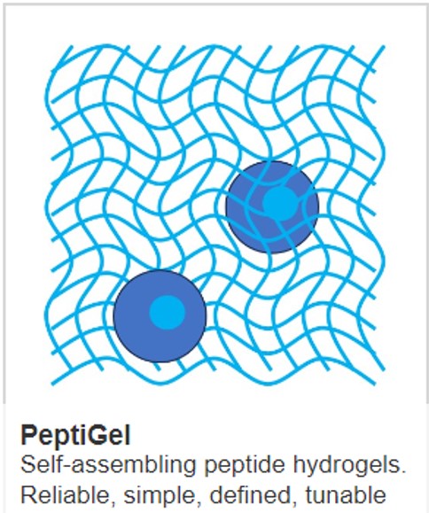
The use of peptide hydrogels for cancer research
Here at Manchester Biogel, we are redefining cell culture through our PeptiGels (fully synthetic peptide hydrogels) – which have already been proven successful in supporting a wide range of cancer applications, such as:
- The use of the extracellular matrix (ECM) in the study of Pancreatic Cancer
- The use of the extracellular matrix (ECM) in the study of Breast Cancer
- Development of PeptiGel-based 3D in vitro Model of Epithelial Ovarian Cancer
The core technology benefits of our PeptiGels include:
- functionally and mechanically fine-tuning to create an in vitro environment that mimics the in vivo and essentially any human tissue
- they are supplied ready-to-use
- they are chemically defined
- there is no batch-to-batch variability, which gives researchers reliable and reproducible results while saving time and resource
IMAGE: Regemat 3D print head. Credit: CellGS
Learn more about powerful technologies that are enabling research: