MISEV: extracellular vesicle research standards

A shared understating of data and its interpretation is fundamental to science. In biological systems, complexity and variability must be communicated in a way that can be understood and interpreted by other labs. To facilitate scientific communication of data between labs working on extracellular vesicles (EVs), a common language of data standards has been developed.
What is MISEV?
The International Society for Extracellular Vesicles (ISEV) has published and twice updated a set of guidelines for researchers in EV studies, known as "Minimal information for studies of extracellular vesicles". The latest set is MISEV2023. This framework covers good practices, up-to-date isolation and characterization methods and defines the information required for new studies in the EV field.
Good practices for EV research
As EV research expands, so does the need for standardized reporting of new data. The guidelines set out in MISEV are designed to ensure rigorous data collection and reproducibility.
This provides a number of recommended techniques for concise reporting on EV studies to demonstrate the presence of particular EV sub-types. This includes:
Quantification of particle size
Measuring EV populations in terms of size and particle concentration relies on the use of instruments designed for viewing nano-sized particles, such as NTA instruments and flow-cytometry. This is important, as it allows the researcher to identify the different populations of vesicles based on their size, as well as confirm whether a purification technique has yielded the right particle size. These metrics can be used in downstream applications, for example, to estimate how many vesicles are needed to exert an effect on target cells.
Protein quantification
The total protein within an EV preparation can be investigated using a number of different assays and techniques. To avoid false readings from co-purified non-EV proteins, the protein concentration should only be used as an EV readout when working with highly pure EV isolates. Moreover, protein concentration alone should be avoided when quantifying EVs, but used in conjunction with multiple other techniques. It should also be reported whether intact or disrupted EV preparations are used with any protein quantification technique.
Total lipid quantification
In recent years, more researchers are investigating lipid compositions of EV preparations and the role different lipid types play in EV biology. Colourimetric assays are a useful tool in quantifying total lipid concentrations, but it should be considered whether contaminating lipids such as lipoproteins are also being measured.
EV protein composition
Due to the heterogeneity of EVs, it can be difficult to understand which protein markers to measure when trying to characterise EV preparations. To address this, a framework set out by MISEV recommends different categories of protein markers that can help identify both the presence of specific EV populations, as well as the purity and the possible intracellular origins. These proteins are divided up into five categories. Categories 1 and 2 covers conserved proteins found across most, if not all EV populations, and includes the tetraspanin markers CD9, CD81 and CD63, as well as cytosolic markers such as ALIX and TSG101). Category 3 proteins are used to assess the purity of preparations and include lipoprotein markers such as apoA1 and ApoB, as well as immunoglobulins. Categories 4 and 5 proteins provide further information on the intracellular origins of the EVs, as well as any co-isolates in the EV preparations. These markers can include histones and cytochrome C for intracellular origin, or cytokines such as IFN-y for co-isolates.
Future directions
As research into EVs continues, so does the expanding need for standardization. The guidelines set out by MISEV condense a plethora of knowledge from numerous experts in the field. By following the techniques explained here, innovative research can continue with increased reproducibility and rigour.
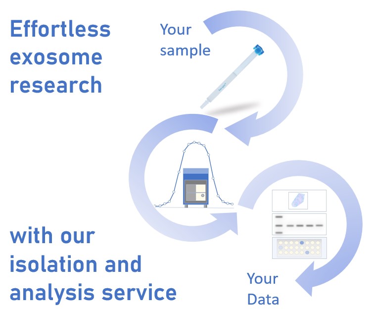
Cell Guidance Systems helps researchers, both new and experienced in the EV field, by providing qualified reagents and EV Isolation and Analysis services that enable researchers to provide their sample of interest and receive highly pure EVs, as well as a detailed report that covers all of the characterisation techniques outlined in MISEV. Utilising dedicated instruments and our widely cited Exo-spin technology, Cell Guidance Systems can help accelerate your research with confidence.
IMAGE - Exosome CREIDT Guillaume Pelletier
#/media/File:Exosome_with_hsp70.png)
Learn more about powerful technologies that are enabling research: