Under pressure: Modeling glaucoma using sustained cytokine release PODS

Primary open-angle glaucoma (POAG) remains the most common cause of irreversible blindness worldwide. Central to its pathogenesis is dysfunction of the trabecular meshwork (TM), a specialized collagenous tissue located at the iridocorneal angle of the eye. TM cells (TMCs) actively remodel the extracellular matrix (ECM) to maintain intraocular pressure (IOP) homeostasis. In POAG, aberrant ECM deposition, stiffening, and fibrotic remodeling impair fluid outflow, leading to elevated IOP and progressive vision loss.
Developing robust in vitro models of the TM is therefore essential for advancing both mechanistic understanding and therapeutic discovery. However, current culture systems rarely capture the architectural complexity of the TM or the subtle interplay of biophysical and biochemical cues that govern TMC behavior.
A study from the University of Birmingham published in Acta Biomaterialia overcomes this barrier by integrating biomimetic collagen scaffolds with Polyhedrin Delivery Systems (PODS®), enabling controlled delivery of transforming growth factor-beta 2 (TGF-β2), a cytokine central to glaucoma pathology.
PODS: Sustained and Localized Protein Release
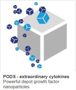
PODS are protein microcrystals that encapsulate biologically active molecules. Their key advantage lies in sustained, spatially restricted release of encapsulated factors. Unlike conventional soluble supplementation, where proteins degrade rapidly and require repeated dosing, PODS release bioactive proteins steadily over time, creating a chronic exposure profile that more closely mimics in vivo conditions. In the context of glaucoma, this is particularly relevant.
Engineering a Pathophysiological Microenvironment
The researchers first recreated the structural properties of the juxtacanalicular tissue (JCT), the TM’s innermost layer and the primary site of outflow resistance. Using confined plastic compression (applying pressure to a material within a mold), they increased collagen fiber density and anisotropy, producing a scaffold with reduced pore size and enhanced stiffness, closely resembling the native JCT. TMCs embedded within this compressed collagen displayed hallmark features of a partial mesenchymal state, including cytoskeletal reorganization and elastin deposition.
To model disease progression, PODS-TGF-β2 was incorporated into these scaffolds. This addition induced a robust mesenchymal phenotype in TMCs, characterized by increased α-smooth muscle actin, fibronectin, and enhanced contractility. Gene expression analyses revealed coordinated upregulation of mesenchymal transcription factors such as TWIST1 and N-cadherin, mirroring profiles observed in glaucomatous tissue. Importantly, these changes were only consistently observed with PODS-mediated cytokine delivery, which maintained local, physiologically relevant exposure over several days.
Utility of PODS in Biomedical Research
The study demonstrates several key utilities of PODS technology:
- Physiological relevance – By providing prolonged cytokine release, PODS recreate the chronic exposure patterns that underlie fibrosis in glaucoma, avoiding the non-physiological surges typical of bolus dosing.
- Spatial fidelity – Embedding PODS within collagen matrices permits local release, enabling gradient formation and microenvironmental control that are otherwise difficult to achieve.
- Experimental reproducibility – PODS protect encapsulated proteins from degradation, ensuring stable concentrations across replicates and reducing batch variability.
- Versatility – While this study focused on TGF-β2, PODS can encapsulate a wide range of proteins, offering a modular platform to interrogate diverse signaling pathways.
Broader Implications
The integration of PODS-derived TGF-β2 into 3D TM models provides an unprecedented tool for studying POAG pathogenesis. It enables researchers to dissect how ECM architecture and sustained cytokine signaling interact to drive TMC plasticity and fibrotic remodeling. Moreover, it establishes a predictive platform for pre-clinical drug screening, where candidate therapeutics can be evaluated against physiologically relevant disease states rather than oversimplified monolayers.
Beyond glaucoma, the implications of PODS technology extend widely. Controlled protein delivery is central to tissue engineering, regenerative medicine, fibrosis research, and oncology. By enabling precise manipulation of cellular environments, PODS facilitate the study of complex biological processes that unfold over days to weeks, bridging the gap between static culture models and the dynamic physiology of living tissues.
This study positions PODS as a transformative tool for disease modeling. By enabling sustained, localized delivery of TGF-β2, PODS not only illuminate the mechanisms driving trabecular meshwork dysfunction in glaucoma but also establish a generalizable strategy for creating more faithful in vitro models. As research increasingly demands systems that replicate the intricacy of human disease, PODS technology offers both the precision and versatility needed to meet that challenge.
IMAGE: Trabecular meshwork CREDIT: Wikimedia commons
Learn more about powerful technologies that are enabling research:


