Using PeptiInk® Alpha-1™ for bioprinting chondrocytes

The complex structure of articular cartilage and its lack of vasculature present particular challenges for regenerative medicine. A recent study led by researchers at University College London explored the utility of PeptiInk® Alpha 1™ for printing 3D chondrocyte structures.
Damage to articular cartilage can lead to osteoarthritis, a disease that is widespread in older people in Western countries. As well as the huge medical cost and pain endured, loss of mobility in the individuals impacts their ability to contribute to society. At present, the early stages of the disease are primarily treated with pain killers and later stage disease is subject to joint replacement surgery. Interventions that provide viable alternatives to slow or even prevent the progress of osteoarthritis are needed.
One of the challenges in treating cartilage is its complex structure and lack of vasculature. In order to generate complex cartilage structures for transplant, there is widespread interest in the use of 3D bioprinting whereby cells suspended in hydrogels are printed in spatially defined structures using 3D computer-aided design (3D CAD). The hydrogel may be deposited with various printing technologies including extrusion, jet and vat photopolymerization. Most researchers use extrusion.
There are a number of considerations for the choice of hydrogel. One of the most important considerations is the hydrogels’ shear-thinning properties. Shear-thinning is a non-Newtonian property of some hydrogels which lose viscosity when under shear stress (i.e. when they are being disrupted/squeezed). Shear-thinning enables the extrusion and pipetting of hydrogels. Other important hydrogel characteristics include biodegradability (to allow invasion of cells and matrix replacement by matrices secreted by the cells) and biocompatibility.
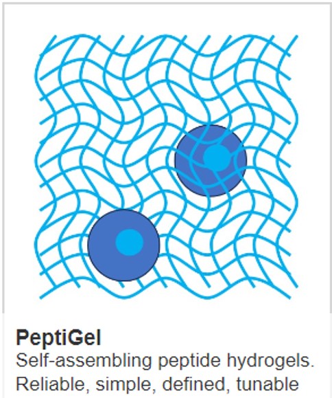
The most widely used materials for printing are complex, alginate-based. However, these hydrogels are not easy to scale and have sub-optimal mechanical properties. PeptInk Alpha 1 represents a distinct improvement on alginate and was selected for assessment by the research team. PeptiInks are self-assembling peptide-based hydrogels. They are readily extruded and their elastic modulus increases on contact with culture medium which changes the hydrogel’s pH and introduces ions to promote further hydrogel cross-linking.
Human primary chondrocytes were printed and assessed, over a period of 14 days, with a range of standard assays for cell viability and markers of chondrogenesis. Cell viability gradually increased over time with cells exceeding 60%, much higher than can be achieved with the gold standard cell pellet which develops a necrotic core. Chondrocyte differentiation was assessed with a range of markers. Early chondrogenesis was demonstrated by strong expression of SOX-9. Later, expression of collagen type II and aggrecan were also shown to increase.
The study demonstrates the benefits of using PeptiInk Alpha 1 for bioprinting. In particular, the study showed that Alpha 1 enabled chondrocytes to survive and also promoted chondrogenesis.
IMAGE: Aggrecan (a marker of chondrogenesis) and DAPI (a nuclear stain) stained chondrocytes on day 14 following printing in PeptiInk Alpha 1. CREDIT Sants-Beato et al
Learn more about powerful technologies that are enabling research: