How to generate microRNA sequence data from EVs

Introduction
Extracellular vesicles (EVs), including exosomes, are lipid-membrane bound, cell-derived nanoparticles secreted by all cell types under physiological and pathological conditions. They are found in most biological fluids including plasma, serum, urine, saliva, and cerebrospinal fluid (CSF). EV transport packages of proteins, lipids, and nucleic acids including microRNAs that reflect the physiological state of their cell of origin. As a result, EVs have emerged as powerful vehicles for biomarker discovery and liquid biopsy.

Sample input material
This workflow works with a range of biological samples including plasma, serum, saliva, urine, cerebrospinal fluid (CSF), and cell culture media.
Step 1. Purification of EVs with Exo-spin™ Size Exclusion Chromatography (SEC)
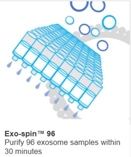
Exo-Spin™ from Cell Guidance Systems utilizes SEC which separates EVs by size using porous beads to separate EVs. Exo-spin™ preserves EV structure and efficiently separates them from soluble proteins. Exo-spin™ kits have been used in hundreds of EV studies and have shown consistent results for small RNA profiling.
Input volume: 0.5 to 1 mL of plasma/serum; up to 5 mL of CSF.
Typical yield: >109 EV particles; 20–100 ng RNA.
Step 2 isolate microRNA from the EVs
A variety of kits are available to extract RNA. Trizol is a simple solution that in our hands, and other groups, performs better for RNA extraction from EVs than affinity columns.
Step 3 Amplify the microRNA
NEXTFLEX from Revvity yields excellent results. In particular, NEXTFLEX Small RNA Sequencing Library Prep Kit V4 is specifically designed for enriching and sequencing small RNAs, with features like blood miRNA blockers to remove common blood-specific miRNAs, improving data analysis.
Step 4 Perform sequencing reaction and sequence generation
A variety of platforms from manufacturers including Illumina, such as MiSeq, have been used for this step.
Step 5 Analysis
A variety of web-based tools are available for analysis of mIRNA. Some of the best tools include TargetScan for target prediction, miRTarBase and DIANA-TarBase for experimentally validated miRNA-target interactions, and miRNet for network analysis and visualization.
References
- Indication of horizontal DNA gene transfer by extracellular vesicles
- Comprehensive analyses of miRNAs revealed miR-92b-3p, miR-182-5p and miR-183-5p as potential novel biomarkers in melanoma-derived extracellular vesicles
IMAGE: RNA in exosomes CREDIT: Shutterstock
Learn more about powerful technologies that are enabling research:


