Growth factors for organoid production

Organoids have moved in little more than a decade from a curiosity to a central tool of modern cell biology. These self-organising, three-dimensional miniature tissues recapitulate the architecture and at least some of the function of their parent organs, and they have become indispensable for studying development, modelling disease, screening drugs, and exploring regenerative therapies. Yet behind every successful organoid culture lies an often-underappreciated dependency: a precisely choreographed cocktail of growth factors. The cells that build an organoid do not self-organise in a vacuum. They require the same signalling cues that orchestrate their behaviour in the living body, supplied in the right combination, at the right concentration, and over the right timescale.
This article surveys the growth factors that underpin organoid culture, why they matter, and how their delivery shapes organoid quality, reproducibility and cost. It explains the roles of the core signalling families, why stem-cell niches depend on tightly regulated factor gradients, and how the practical challenges of growth factor instability and expense are driving interest in sustained-release approaches. For researchers establishing or scaling organoid work, understanding the logic of these signalling requirements is the difference between robust, reproducible cultures and frustrating, variable ones.
| Key Point |
| Organoids depend on growth factors to recreate the signalling niche of their parent tissue. The core factors fall into a handful of conserved families: the Wnt and R-spondin axis sustains stem-cell self-renewal; EGF drives proliferation; FGFs pattern and expand tissues; BMPs and their antagonists (Noggin, Gremlin) balance differentiation against stemness; and TGF-beta family members tune the whole system. Because most of these proteins are expensive, unstable in culture medium, and require frequent replenishment, growth factor supply is one of the largest practical and financial constraints on organoid work. Sustained-release formats that deliver factors at physiological concentrations over days, rather than as repeated bolus additions, offer a route to more reproducible cultures at lower cost. |
Why Organoids Need Growth Factors
In a living organ, stem and progenitor cells reside in a niche: a specialised microenvironment that supplies the signals controlling whether a cell stays quiescent, divides, or differentiates. The niche is built from neighbouring cells, extracellular matrix, and a constellation of secreted signalling proteins. Remove a stem cell from this niche and culture it on plastic in simple medium, and it rapidly loses its identity, either differentiating uncontrollably or dying.
Organoid culture is, in essence, an attempt to rebuild the niche in vitro. A supportive matrix (most commonly a basement-membrane extract or a defined synthetic hydrogel) provides the physical scaffold and adhesion cues, while the culture medium supplies the soluble signals. The growth factors in that medium are not generic "feed" for the cells; they are specific instructions. Each one engages a receptor and triggers a signalling cascade that the cell interprets as a command to proliferate, survive, migrate, or commit to a particular fate. Get the combination wrong, and the culture fails to form proper structures, drifts toward the wrong cell types, or cannot be maintained beyond a few passages.
The remarkable insight from the foundational organoid work of the early 2010s was that a relatively small set of factors, supplied in the right balance, is sufficient to recapitulate the essential niche signals for many tissues. Identifying that minimal set for each organ type, and refining the concentrations, remains an active area of method development.
The Wnt and R-spondin Axis: Maintaining Stemness
For organoids derived from many epithelial tissues, particularly the intestine, stomach, liver and pancreas, the Wnt signalling pathway is the master regulator of stem-cell self-renewal. Wnt proteins bind Frizzled receptors and stabilise beta-catenin, which then drives transcription of genes that keep cells in a proliferative, undifferentiated state. In the intestinal crypt, a Wnt gradient defines the stem-cell zone: high Wnt at the crypt base maintains stemness, while declining Wnt up the crypt-villus axis permits differentiation.
R-spondin acts as a crucial amplifier of Wnt signalling. It binds LGR receptors (LGR4, LGR5 and LGR6) and neutralises the membrane enzymes RNF43 and ZNRF3 that would otherwise degrade Frizzled receptors. The result is that R-spondin sensitises cells to whatever Wnt is present, sharply boosting the pathway. LGR5 is itself the canonical marker of adult epithelial stem cells, which makes R-spondin responsiveness almost synonymous with stemness in these tissues. In practice, R-spondin is one of the most important, and most expensive, components of intestinal and gastric organoid media.
Wnt proteins themselves are notoriously difficult to handle. They are lipid-modified, poorly soluble, and lose activity rapidly once diluted into aqueous medium. Many laboratories rely on Wnt-conditioned medium produced from engineered cell lines rather than purified protein, which introduces batch-to-batch variability that is a recognised source of irreproducibility in organoid research.
EGF: The Proliferative Workhorse
Epidermal growth factor (EGF) is the most ubiquitous growth factor in organoid media, appearing in protocols for intestine, stomach, liver, pancreas, lung, mammary tissue and more. EGF binds the EGFR receptor tyrosine kinase and activates the MAPK and PI3K pathways, driving cell-cycle entry and proliferation. Where Wnt and R-spondin maintain the stem-cell state, EGF supplies much of the mitogenic push that expands the organoid.
EGF is comparatively robust and inexpensive relative to Wnt or R-spondin, but it is not without subtlety. Excess EGF can override differentiation signals and bias cultures toward an undifferentiated, hyperproliferative phenotype, while too little limits growth. The concentration must be balanced against the other factors in the medium, and in some differentiation protocols EGF is deliberately withdrawn to allow cells to mature.
FGFs: Patterning and Expansion
The fibroblast growth factor family comprises more than twenty members with diverse, tissue-specific roles. In organoid culture, particular FGFs are selected to match the requirements of the target tissue. FGF2 (basic FGF) is a broadly used mitogen and survival factor. FGF7 (keratinocyte growth factor) and FGF10 are central to branching morphogenesis and epithelial expansion in lung, pancreas, salivary gland and mammary organoids, where they signal from the surrounding mesenchyme to the epithelium. FGF4 features in protocols for liver and intestinal patterning.
FGFs typically signal through FGFR receptor tyrosine kinases and, like EGF, drive proliferation via MAPK and PI3K. Their tissue specificity comes both from which FGF is supplied and from which FGFR isoforms the target cells express. Many FGFs require heparin or heparan sulfate as a co-factor for stable, high-affinity receptor binding, which is one reason FGF activity in culture can be inconsistent if this requirement is not met. FGF2 in particular is thermally unstable and loses much of its activity within a day at 37 degrees Celsius, a property that has direct consequences for how often medium must be refreshed.
BMPs and Their Antagonists: The Differentiation Balance
Bone morphogenetic proteins (BMPs), members of the TGF-beta superfamily, generally promote differentiation and oppose stem-cell expansion in epithelial organoids. This makes them a double-edged tool. In the intestinal crypt, BMP signalling is high toward the villus tip and low at the crypt base, and this gradient helps drive the transition from stem cell to differentiated enterocyte.
To maintain stemness and expand organoids, protocols therefore frequently include BMP antagonists rather than BMPs themselves. Noggin is the most widely used: it binds BMP ligands and prevents them from engaging their receptors, thereby protecting the stem-cell compartment. Gremlin 1 is another BMP antagonist used in some protocols. The interplay is delicate: enough BMP inhibition to preserve stemness during expansion, then controlled restoration of BMP signalling (by withdrawing Noggin or adding BMP) to drive differentiation when mature cell types are desired. This switchable control over the BMP axis is one of the clearest examples of how organoid fate is steered by manipulating growth factor balance over time.
| Why Timing and Balance Matter |
| Organoid protocols usually divide into an expansion phase and a differentiation phase, and the growth factor recipe differs sharply between them. Expansion media are rich in stemness-promoting factors (Wnt, R-spondin, EGF) and BMP antagonists (Noggin), keeping cells proliferative and undifferentiated. Differentiation media withdraw or reduce these and may add factors that push cells toward specific fates. The transition is not simply on or off; the relative concentrations of competing signals at any moment determine the proportion of stem, progenitor and mature cells in the organoid. Because each factor decays at its own rate in the medium, the effective balance drifts continuously between feeds, which is a major and often unappreciated source of variability. Delivery systems that hold factor concentrations steady would reduce this drift and make the differentiation trajectory more controllable. |
TGF-beta, Nodal, Activin and Other Modulators
Beyond the core quartet, a range of additional factors fine-tunes organoid behaviour. The broader TGF-beta family, including TGF-beta itself, Activin and Nodal, plays context-dependent roles. In pluripotent stem cell-derived organoids, Activin/Nodal signalling is critical for specifying definitive endoderm, the starting point for gut, liver, lung and pancreatic organoids. In adult epithelial organoids, TGF-beta signalling is often inhibited (using small-molecule inhibitors such as A83-01) because it tends to suppress proliferation and induce differentiation or apoptosis.
Hepatocyte growth factor (HGF) supports liver and several other organoid types, promoting survival and proliferation of epithelial cells. Vascular endothelial growth factor (VEGF) is increasingly important in efforts to vascularise organoids, a major frontier for growing larger and more physiologically realistic constructs. Insulin and IGF-1 support general survival and metabolism and appear in many formulations. Small molecules frequently accompany the protein factors: Y-27632 (a ROCK inhibitor) improves single-cell survival during seeding, while inhibitors of GSK3, TGF-beta and other pathways are used to mimic or block specific signals more cheaply and stably than recombinant proteins can.
Comparing the Core Organoid Growth Factors
| Core Growth Factors in Organoid Culture | ||||
| Factor | Primary Role | Pathway | Common Organoid Types | Stability / Cost |
| Wnt3a | Stem-cell self-renewal | Wnt / beta-catenin | Intestine, stomach, liver | Very unstable; expensive |
| R-spondin | Amplifies Wnt; supports LGR5+ cells | Wnt (via LGR4/5/6) | Intestine, stomach, pancreas | Moderate stability; very expensive |
| EGF | Proliferation / mitogen | EGFR / MAPK, PI3K | Nearly all epithelial | Robust; relatively cheap |
| FGF2 / FGF7 / FGF10 | Expansion, branching, patterning | FGFR / MAPK, PI3K | Lung, pancreas, mammary, liver | FGF2 thermally unstable; moderate cost |
| Noggin | BMP antagonist; preserves stemness | Blocks BMP / SMAD | Intestine, stomach, others | Moderate stability; expensive |
| BMP-2 / BMP-4 | Drives differentiation | BMP / SMAD | Used in differentiation phases | Moderate; moderate cost |
| HGF | Survival, proliferation | MET / MAPK, PI3K | Liver, kidney, others | Moderate stability; moderate cost |
| VEGF | Vascularisation | VEGFR | Vascularised / complex organoids | Moderate; moderate cost |
| Activin A / Nodal | Endoderm specification (PSC-derived) | TGF-beta / SMAD | Gut, liver, lung, pancreas (from PSCs) | Moderate; moderate cost |
The Practical Problem: Instability and Cost
The signalling biology of organoids is elegant, but the practical reality of supplying these factors is anything but. Two interlinked problems dominate: instability and cost.
Most growth factors are recombinant proteins with limited stability in aqueous medium at 37 degrees Celsius. FGF2 is the textbook case, losing the majority of its activity within a day, but EGF, Wnt and many others also degrade over time. Because of this, organoid media must be refreshed every two to three days, with full growth factor supplementation each time. The effective concentration a cell experiences is therefore a sawtooth: a peak immediately after feeding that decays toward a trough before the next change. Cells never see the steady, physiological concentration that the niche provides in vivo. This instability not only wastes expensive protein but also introduces variability, since the rate of decay depends on temperature, handling and medium composition.
The cost problem follows directly. Wnt and R-spondin in particular are among the most expensive reagents in cell biology, and a single organoid experiment maintained over weeks can consume substantial quantities. For laboratories scaling up to high-throughput drug screening or biobanking, growth factor cost can become the dominant line item, sometimes pricing organoid approaches out of reach. The combination of frequent feeding (driven by instability) and high unit cost compounds the burden.
These pressures explain why the field has invested heavily in workarounds: conditioned media from engineered producer cells, small-molecule pathway agonists and antagonists that are cheaper and more stable than proteins, engineered "surrogate" Wnt and R-spondin proteins with improved properties, and, increasingly, sustained-release delivery systems.
Sustained Release: Matching Delivery to the Niche
If the central problem is that bolus addition of unstable proteins produces a fluctuating, wasteful concentration profile, the logical solution is to deliver growth factors in a sustained, controlled manner that holds them near physiological levels over the days between feeds. This is precisely the rationale that has driven sustained-release growth factor technology in regenerative medicine, and the same logic applies to organoid culture.
A depot that releases a growth factor slowly within the culture, or embedded in the matrix surrounding the organoids, would smooth out the sawtooth, maintain a steadier signal, reduce the frequency of feeding, and lower the total amount of expensive protein consumed. It would also protect the encapsulated factor from degradation until the moment of release, addressing the instability problem at source. For factors such as FGF2 that decay rapidly, a system that shields the protein and releases active material gradually is especially valuable.
Cell Guidance Systems' PODS technology embodies this approach. PODS (POlyhedrin Delivery System) crystals encapsulate a growth factor within a stable protein co-crystal and release it gradually under the action of cellular proteases, delivering active protein at sustained, physiological concentrations rather than as a single fluctuating dose. Because the cargo is protected within the crystal lattice until release, PODS-formatted factors retain activity far longer than free protein in medium, and a single application can supply a culture over an extended period. Importantly, release is driven by the proteases that cells themselves secrete, so growth factor is liberated in response to the culture's own activity. For organoid workflows, PODS crystals can be deposited on the culture surface, embedded within the hydrogel matrix, or held separate from the cells in a transwell insert, embedding the growth factor signal within the niche itself rather than relying on repeated bolus addition to the bulk medium.
PODS in Practice: Published Organoid Evidence

PODS crystals under scanning electron microscopy. Each cubic protein co-crystal encapsulates a growth factor and releases it gradually under the action of cellular proteases. Image: Cell Guidance Systems.
The benefit for organoid culture is not merely theoretical, and recent peer-reviewed work makes the case concretely. In a retinal ganglion cell (RGC) organoid model, stem cell-derived organoids were maintained with PODS BDNF and PODS GDNF added as a single application. The published comparison is striking: achieving a similar effect with conventional soluble BDNF and GDNF required adding at least 250 ng of each factor five separate times over the culture period, whereas one dose of PODS sufficed. The PODS-treated organoids were not merely equivalent but better, showing smoother-surfaced, healthier morphology and a significant increase in RGC yield across multiple markers, with subtype yields raised up to two-fold. ELISA confirmed steady growth factor release in the region of 50 pg/mL sustained over a week from that single application.
The 2025 follow-up study extended these findings to human stem cell-derived RGCs and to function, not just yield. Organoids supplemented with PODS BDNF/GDNF roughly doubled neurite outgrowth (around 1050 micrometres versus 520 in untreated controls) and showed higher spontaneous firing rates on multielectrode arrays, indicating more mature, functional neurons. When the PODS-treated cells were transplanted, donor survival improved 2.7-fold for mouse RGCs and 15-fold for human RGCs, and the sustained release also protected host neurons. For organoid researchers the message is direct: a single, defined PODS application produced organoids of higher quality and greater functional maturity than repeated bolus dosing, while removing several feeding interventions from the protocol.
The same principle has been validated in a second neuronal organoid system. In stem cell-derived otic (inner-ear) neuronal cultures, PODS BDNF supplied a stable, localised growth factor gradient in a microfluidic device, driving survival, differentiation toward spiral ganglion neurons, and directional neurite outgrowth that a homogeneous bolus of soluble BDNF could not reproduce. Across both retinal and otic models, the recurring observation is that sustained, protease-driven release from PODS recreates the steady, spatially organised signalling that a developing organoid niche actually requires.
Beyond these published case studies, separate long-term release data show detectable growth factor for at least nine weeks following a single PODS application, allowing media changes to be reduced to roughly once a week without replenishing growth factor. For organoid work, where cultures run for weeks and every medium change is an opportunity for variability and contamination, this combination of fewer interventions, steadier signalling and lower total protein consumption directly addresses the instability and cost problems described above. PODS crystals can be incorporated into hydrogels such as PeptiGel or nanofibrillar cellulose, embedded in 3D-printed scaffolds, deposited on the culture surface, or held separate from the cells in a transwell insert, giving researchers flexible control over where and how the signal is delivered within the organoid niche.
Toward More Reproducible Organoid Culture
Reproducibility is the recurring challenge in organoid research, and growth factor supply sits at its heart. Conditioned media vary between batches; protein lots vary in activity; decay rates vary with handling; and the fluctuating concentrations between feeds mean no two cultures experience exactly the same signalling history. Standardising growth factor delivery, whether through defined recombinant proteins, surrogate ligands, or sustained-release formats such as PODS, is one of the most direct routes to more reliable organoids. By holding factor concentrations steady from a single defined application and removing several handling steps from the protocol, sustained release reduces two of the largest sources of run-to-run variation at once.
As organoids move from research tools toward applications in personalised medicine, drug development and even transplantation, these considerations only grow in importance. Regulatory and clinical contexts demand defined, reproducible, animal-product-free culture systems. The trend is clearly toward fully defined media, synthetic matrices in place of animal-derived basement membrane extracts, and controlled, sustained delivery of the signalling proteins that make organoids possible.
Summary
Growth factors are not an accessory to organoid culture; they are its foundation. The Wnt and R-spondin axis sustains the stem-cell compartment, EGF and FGFs drive proliferation and patterning, and the BMP and TGF-beta families, together with their antagonists, balance self-renewal against differentiation. Supplying this signalling cocktail in the right combination, concentration and sequence is what allows a handful of cells to self-organise into a functional miniature tissue.
The chief obstacles are practical: most of these proteins are unstable in culture and expensive to buy, forcing frequent, costly feeding and introducing variability that undermines reproducibility. Sustained-release delivery, which holds growth factors at steady physiological concentrations and protects them from degradation, offers a compelling answer, reducing cost and improving consistency. As organoid technology matures and moves toward clinical and high-throughput applications, controlled growth factor delivery is set to become a defining feature of robust, reproducible culture.
Cell Guidance Systems Growth Factor Products and Services
PODS Growth Factors. For sustained, controlled delivery in organoid culture, our PODS growth factors encapsulate bioactive proteins in a stable protein co-crystal that releases active factor over weeks from a single application. A single dose can replace repeated growth factor additions, cutting feeding frequency, improving consistency between wells and experiments, and lowering the total quantity of expensive protein consumed. The range includes PODS-formatted FGF, BDNF, GDNF, BMP-2, VEGF, Activin and many other factors relevant to stem-cell and organoid work, in both human and other-species formats.
PODS Technology and Resources. For application notes, technical guides and peer-reviewed publications on using PODS in 3D and organoid culture, including the retinal ganglion organoid and long-term release studies referenced above, see our PODS resources page and the PODS depot growth factors technology overview.
Standard Growth Factors. Cell Guidance Systems also offers a comprehensive range of conventional recombinant growth factors for cell and organoid culture.
Custom PODS Proteins. For growth factors outside our standard catalogue, the Custom PODS Proteins service develops sustained-release formulations of your protein of interest.
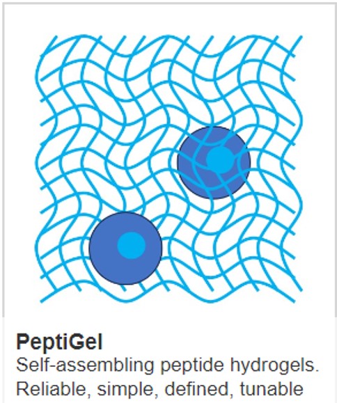
PeptiGel Biomaterials. Our PeptiGel synthetic peptide hydrogels provide a chemically defined, animal-free 3D matrix for organoid culture that can be functionalised with PODS growth factors for niche-embedded, sustained delivery.
References
[1] Sato T, Vries RG, Snippert HJ, et al. Single Lgr5 stem cells build crypt-villus structures in vitro without a mesenchymal niche. Nature. 2009;459(7244):262-265.
[2] Clevers H. Modeling development and disease with organoids. Cell. 2016;165(7):1586-1597.
[3] Sato T, Stange DE, Ferrante M, et al. Long-term expansion of epithelial organoids from human colon, adenoma, adenocarcinoma, and Barrett's epithelium. Gastroenterology. 2011;141(5):1762-1772.
[4] de Lau W, Barker N, Low TY, et al. Lgr5 homologues associate with Wnt receptors and mediate R-spondin signalling. Nature. 2011;476(7360):293-297.
[5] Lancaster MA, Knoblich JA. Organogenesis in a dish: modeling development and disease using organoid technologies. Science. 2014;345(6194):1247125.
[6] Spence JR, Mayhew CN, Rankin SA, et al. Directed differentiation of human pluripotent stem cells into intestinal tissue in vitro. Nature. 2011;470(7332):105-109.
[7] Janda CY, Dang LT, You C, et al. Surrogate Wnt agonists that phenocopy canonical Wnt and beta-catenin signalling. Nature. 2017;545(7653):234-237.
[8] Fujii M, Matano M, Toshimitsu K, et al. Human intestinal organoids maintain self-renewal capacity and cellular diversity in niche-inspired culture condition. Cell Stem Cell. 2018;23(6):787-793.
[9] Hofer M, Lutolf MP. Engineering organoids. Nature Reviews Materials. 2021;6(5):402-420.
[10] Kratochvil MJ, Seymour AJ, Li TL, et al. Engineered materials for organoid systems. Nature Reviews Materials. 2019;4(9):606-622.
[11] Soucy JR, Oswald J, Kriukov E, et al. Sustained neurotrophic factor co-treatment enhances donor and host retinal ganglion cell survival in mice. Translational Vision Science & Technology. 2025;14(9):27.
[12] Chang H, Heuer R, Oleksijew A, et al. An engineered three-dimensional stem cell niche in the inner ear by applying a nanofibrillar cellulose hydrogel with a sustained-release neurotrophic factor delivery system. Acta Biomaterialia. 2020;108:111-127.
[13] Nella KT, Norton BM, Chang HT, et al. Bridging the electrode-neuron gap: finite element modeling of in vitro neurotrophin gradients to optimize neuroelectronic interfaces in the inner ear. Acta Biomaterialia. 2022;151:360-378.
MAIN IMAGE: From Norton et al, 2023