Hidden talents: Cancer diagnostic EVs

Cancer remains one of the leading causes of mortality worldwide, with an estimated 19.9 million new cases diagnosed globally in 2022 [1]. Despite considerable advances in imaging, tissue biopsy and molecular diagnostics, the reliable detection of cancer at an early, treatable stage remains a fundamental challenge across most tumour types. Exosomes are a type of extracellular vesicle (EV). They have attracted intense research interest as a potential solution: nanoscale vesicles shed constitutively by both normal and tumour cells, carrying molecular cargo that reflects the biology of their cell of origin and accessible through minimally invasive sampling of blood, urine or other biofluids.
This article provides a practical scientific overview of exosome biomarkers in cancer: the biological rationale, the cargo classes under active investigation, the cancer types where the evidence base is most advanced, and the methodological requirements that researchers need to meet to generate reliable, publication-ready data.
For researchers new to exosome biology, our introductory article on EV isolation methods provides useful background: UC vs SEC vs TFF: Exosome and EV Isolation Methods Compared.
Key Point |
|
Tumour-derived exosomes carry proteins, miRNAs and nucleic acids that reflect the mutational and transcriptional state of the tumour. Because they can be isolated from blood or urine, exosomes offer a genuinely non-invasive window into tumour biology that complements tissue biopsy and conventional circulating tumour DNA (ctDNA) approaches. |
Why Exosomes Are Biologically Suited to Cancer Biomarker Discovery
Exosomes are membrane-enclosed vesicles of 30 to 150 nm in diameter, released by cells through the fusion of multivesicular endosomes with the plasma membrane. Their biogenesis through the endosomal pathway means that their interior is enriched with proteins, messenger RNAs, microRNAs (miRNAs) and DNA fragments that were present in the cytoplasm or nucleus of the parent cell at the time of release [2].
Several properties make exosomes particularly well suited to biomarker discovery in cancer:
Tumour-selective cargo. Tumour cells frequently release exosomes at higher rates than their normal counterparts, and the molecular cargo reflects the altered proteomic and transcriptomic landscape of the tumour. Driver mutations, splice variants and oncogene amplifications present in the tumour cell can be detected in exosomal protein and nucleic acid cargo.
Protection from degradation. The lipid bilayer membrane of exosomes protects encapsulated miRNAs and proteins from the nucleases and proteases present in plasma, making exosomal biomarkers considerably more stable ex vivo than free circulating miRNAs or proteins in the same sample.
Biofluid accessibility. Exosomes are present at measurable concentrations in plasma, serum, urine, cerebrospinal fluid, saliva and pleural effusions. Blood sampling is sufficient for most cancer biomarker applications, and the isolation procedure does not require the same technical demands as tissue biopsy or surgical resection.
Functional relevance. Tumour-derived exosomes are not passive bystanders: they actively participate in tumour progression, pre-metastatic niche formation and immune evasion [3]. Biomarkers present in exosomes therefore reflect biologically active processes rather than passive cell shedding, which may increase their relevance as indicators of disease stage and therapeutic response.
Classes of Exosomal Cargo Under Investigation as Cancer Biomarkers
Protein Biomarkers
Exosomal surface proteins are among the most accessible and analytically tractable cancer biomarkers. The tetraspanins CD9, CD63 and CD81 and the ADMA10 protein are generic exosome markers present on virtually all exosomes and serve as quality controls for isolation rather than cancer-specific signals. Cancer-relevant protein biomarkers are typically enriched on tumour-derived exosome subpopulations and require assays capable of distinguishing them from the background of exosomes derived from normal circulating and tissue cells.
Glypican-1 (GPC-1) is one of the most extensively studied tumour-associated exosomal proteins. A landmark study by Melo and colleagues demonstrated that GPC-1-positive exosomes could be detected in the serum of pancreatic cancer patients, including those with early-stage disease, with high sensitivity and specificity compared to healthy controls and patients with benign pancreatic disease [4]. This study was important in demonstrating that exosomal surface proteins could discriminate early cancer from non-malignant conditions, a distinction that is rarely achievable with soluble serum protein markers such as CA19-9.
Other protein biomarkers under investigation include EpCAM for epithelial cancers, HER2 for breast cancer, EGFR and programmed death-ligand 1 (PD-L1) for lung cancer, and prostate-specific membrane antigen (PSMA) for prostate cancer. In each case, the challenge is developing quantitative assays with sufficient sensitivity to detect tumour-derived exosome subpopulations against the large background of non-tumour exosomes in plasma.
MicroRNA Biomarkers
MicroRNAs (miRNAs) are small non-coding RNAs of approximately 22 nucleotides that post-transcriptionally regulate gene expression. Tumour cells selectively package specific miRNAs into exosomes, and these exosomal miRNA profiles are reproducibly altered in cancer patients relative to healthy controls. A foundational study by Taylor and Gercel-Taylor identified that exosomal miRNA signatures in serum could distinguish ovarian cancer patients from healthy controls with a sensitivity and specificity comparable to the matched tumour tissue miRNA profile, demonstrating that exosomal miRNAs carry biologically relevant information about the cell of origin [5].
The miRNAs most consistently reported as elevated in cancer-patient exosomes across different tumour types include miR-21, miR-155, miR-210 and miR-141, though specific panels vary considerably by cancer type and the population studied. The field has moved increasingly towards multi-miRNA panels rather than single-marker approaches, recognising that no single miRNA achieves the specificity required for clinical application across a population.
DNA and Other Nucleic Acid Cargo
Exosomes also carry double-stranded DNA fragments that can include tumour-specific mutations, and in some contexts long non-coding RNAs (lncRNAs) and circular RNAs (circRNAs) with cancer-associated expression patterns. While sequencing-based approaches to exosomal DNA are less mature than the protein and miRNA fields, interest is growing in using exosomal DNA alongside or instead of conventional ctDNA as a liquid biopsy analyte, particularly in settings where ctDNA is present at very low abundance.
Cancer Types with the Most Advanced EV Biomarker Evidence
The exosome biomarker literature is extensive but uneven: certain cancer types have accumulated substantial peer-reviewed evidence while others remain at an early exploratory stage. The table below summarises the current state of the field for the most-studied cancer types.
Exosome Biomarker Evidence by Cancer Type |
|||
| Cancer Type | Cargo Class | Key Biomarkers Investigated | Evidence Stage |
| Pancreatic | Protein, miRNA | GPC-1, miR-196a, miR-217 | Most advanced; early detection data in clinical cohorts |
| Breast | Protein, miRNA | HER2, EpCAM, miR-21, miR-155 | Multiple validated cohort studies; treatment monitoring data emerging |
| Lung | Protein, miRNA, DNA | EGFR (mutant), PD-L1, miR-21, miR-210 | Active; EGFR mutation detection in exosomal DNA validated in multiple studies |
| Colorectal | miRNA, protein | miR-21, miR-92a, EpCAM | Growing cohort data; staging and recurrence monitoring investigated |
| Prostate | Protein, miRNA | PSMA, PSA, miR-141, miR-375 | Active; potential to distinguish aggressive from indolent disease |
| Ovarian | miRNA, protein | miR-21, miR-200 family, EpCAM | Strong early data; landmark miRNA tissue-matching study published [5] |
Why Isolation Quality Determines Biomarker Reliability
A recurring challenge in the exosome biomarker literature is the poor reproducibility of findings across studies. While biological variation between patient cohorts contributes to this, a substantial proportion of the irreproducibility can be traced to inconsistencies in the isolation method. Isolation protocol differences affect which particle populations are captured, how much co-isolated protein contamination is present, and whether the integrity of encapsulated miRNAs is preserved.
The three most commonly used isolation approaches each have distinct implications for biomarker studies:
Ultracentrifugation (UC). The historical reference method. UC pellets all particles above approximately 100 nm at 100,000 x g but also co-pellets protein aggregates and lipoproteins that confound downstream assays. UC is technically demanding, causes mechanical stress to EV membranes, and shows poor interlaboratory reproducibility. For biomarker discovery studies intended for publication, UC alone is increasingly regarded as insufficient.
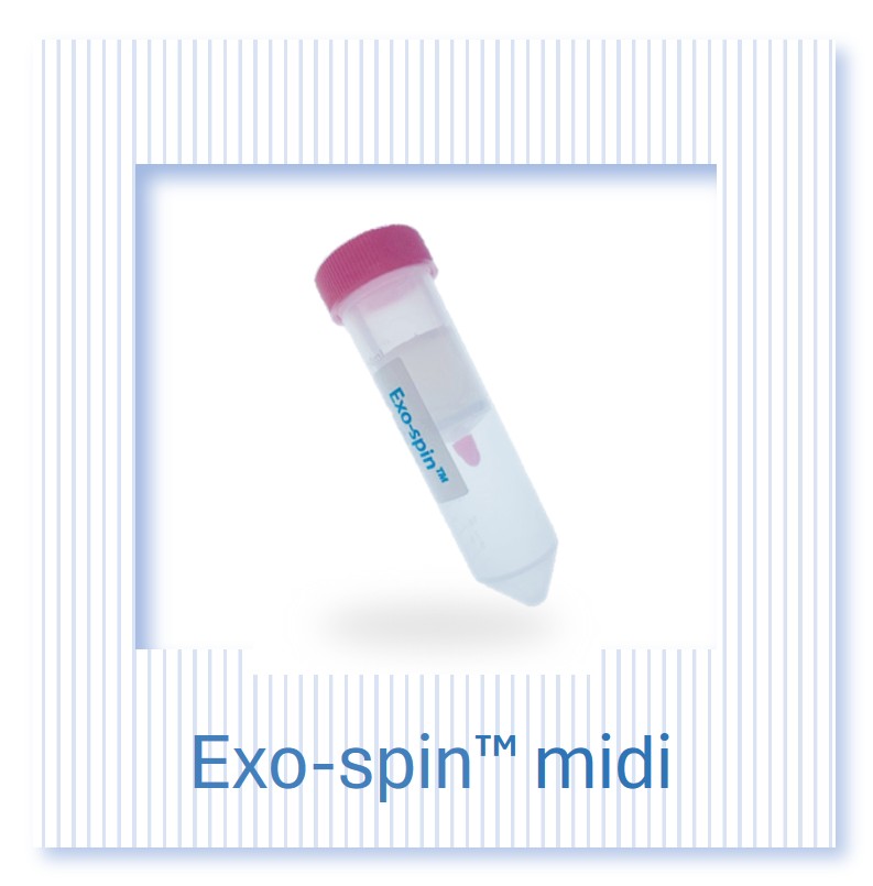
Size exclusion chromatography (SEC). SEC separates EVs from free proteins on the basis of size, providing a purer EV fraction with minimal protein contamination and good preservation of membrane integrity. Exo-spin SEC kits from Cell Guidance Systems provide a rapid, reproducible SEC workflow suitable for plasma and conditioned media volumes of 0.1 to 50 ml. SEC is the method of choice for biomarker studies on small sample volumes where purity is paramount.
Tangential flow filtration (TFF). TFF scales to large conditioned media or biofluid volumes while providing concentration and partial protein depletion in a single continuous run. For biomarker discovery programmes requiring large patient cohort numbers or large starting volumes, TFF upstream of SEC is the most practical approach. Our guide to tangential flow filtration covers the practical workflow in detail.
Practical Guidance |
|
For any cancer biomarker study intended for publication, the isolation method must be fully described and the EV preparation must be characterised using at least particle concentration (NTA), size distribution and tetraspanin expression (CD9, CD63, CD81). Studies relying on precipitation-based isolation kits without purity characterisation are unlikely to meet current peer review standards at major journals. |
Analytical Methods for Exosome Biomarker Characterisation
The analytical toolkit for characterising exosomal biomarkers has expanded considerably over the past decade. The choice of method depends on the cargo class of interest, the required throughput and the downstream application.
Nanoparticle Tracking Analysis (NTA)
NTA quantifies particle concentration and size distribution by tracking the Brownian motion of individual particles in a laser-illuminated field. For cancer biomarker studies, NTA provides the particle count required for normalisation of downstream assays (particles per ml of biofluid) and confirms that the isolation has captured vesicles in the expected size range. Cell Guidance Systems provides a standalone NTA particle analysis service for laboratories that have completed in-house isolation.
Western Blot for Tetraspanin Markers
Western blot detection of CD9, CD63 and CD81 confirms EV identity and provides a semi-quantitative measure of tetraspanin-positive vesicle content. For cancer biomarker studies, tetraspanin western blots serve as positive markers of EV quality, while the absence of contamination markers (calnexin for ER, GM130 for Golgi, albumin for plasma proteins) confirms preparation purity. MISEV2018 guidelines specify both positive and negative marker requirements for publication [2].
ExoLISA Biomarker Assays
ELISA-based quantification of tetraspanin and other surface markers offers a faster, higher-throughput and more quantitative alternative to western blot. Cell Guidance Systems ExoLISA assays provide quantitative detection of CD9, CD63 and CD81, enabling standardised comparisons across samples and time points. In cancer biomarker studies, ExoLISA can be used alongside tumour-marker-specific ELISA assays to generate multi-parameter datasets from the same EV preparation.
MicroRNA Profiling
Exosomal miRNA profiling for cancer biomarker studies typically involves RNA extraction from the EV pellet, followed by quantitative RT-PCR for targeted panels or small RNA sequencing for discovery-phase analysis. A consistent challenge is the small absolute quantity of RNA recoverable from plasma-derived exosomes: rigorous normalisation strategies, technical replicates and the inclusion of appropriate spike-in controls (such as synthetic cel-miR-39) are essential for reliable quantification. Sample-matched normal controls and blinded analysis are minimum requirements for any biomarker discovery study.
Mass Spectrometry for Proteomic Cargo
Label-free quantitative proteomics and targeted mass spectrometry methods are increasingly applied to exosome preparations from cancer patient biofluids. These approaches can simultaneously identify hundreds of exosomal proteins and compare their relative abundance between cancer and control groups. For successful application, the EV preparation must be sufficiently pure: high residual albumin, immunoglobulin or other abundant plasma proteins will suppress detection of lower-abundance EV cargo proteins.
Recommended Characterisation Framework for Cancer Biomarker Studies
MISEV2018 provides the community-agreed minimum reporting standards for EV studies [2]. For cancer biomarker research, the following characterisation framework is recommended as a minimum before data submission to a peer-reviewed journal:
MISEV-Compliant Characterisation Checklist for Cancer Biomarker Studies |
||
| Measurement | Method | Purpose |
| Particle concentration and size | NTA | Confirms vesicle presence, provides normalisation denominator |
| Tetraspanin positive markers | Western blot or ExoLISA (CD9, CD63, CD81) | Confirms EV identity |
| Total protein content | BCA assay | Particle-to-protein ratio; indicates preparation purity |
| Negative markers | Western blot (calnexin, albumin) | Excludes non-EV contamination |
| Morphology | Transmission electron microscopy (where available) | Confirms cup-shaped vesicle morphology; recommended but not always mandatory |
| Isolation method description | Full protocol in methods section | Required for reproducibility and MISEV compliance |
Cell Guidance Systems Products for Cancer Biomarker Research
Exosome Isolation and Characterisation at Cell Guidance Systems |
|
Cell Guidance Systems supplies a complete portfolio of EV isolation and characterisation tools for cancer biomarker research, from small-volume SEC isolation kits to large-scale TFF systems and quantitative biomarker assays. Our outsourced EV services provide MISEV-compliant characterisation for laboratories without in-house analytical capability. |
Exo-spin SEC kits. For plasma, serum or small conditioned media volumes (0.1 to 50 ml), Exo-spin size exclusion chromatography kits provide rapid, high-purity EV isolation with minimal protein contamination. This is the recommended approach for cancer biomarker studies where sample volume is not a limiting factor and purity is the primary consideration.
EVlution TFF system. For large conditioned media volumes or large patient cohort plasma processing programmes, the EVlution TFF system concentrates EVs from up to several litres of input material in a single continuous run. Used upstream of Exo-spin SEC, EVlution provides both scale and purity.
ExoLISA assays. ExoLISA CD9, CD63 and CD81 assays provide quantitative tetraspanin detection from EV preparations, enabling standardised characterisation across sample sets and time points consistent with MISEV requirements.
EV Service Packages. For laboratories without in-house isolation or characterisation equipment, the Cell Guidance Systems EV Basic and EV MISEV Service Packages provide outsourced isolation, NTA analysis, protein quantification, tetraspanin western blot and ExoLISA biomarker detection. Contact our scientific team to discuss your requirements.
Summary: Key Considerations for Exosome Biomarker Studies in Cancer
Exosome Biomarker Study: Key Decisions |
|
| Decision Point | Recommendation |
| Small plasma or serum volume (under 50 ml) | Exo-spin SEC isolation |
| Large patient cohort or high-volume biofluid processing | EVlution TFF followed by Exo-spin SEC |
| Tetraspanin quantification for MISEV compliance | ExoLISA assays (CD9, CD63, CD81) |
| Particle concentration and size distribution | NTA (in-house or via CGS NTA service) |
| No in-house isolation or characterisation capability | CGS EV Service Package |
Exosome biomarkers represent one of the most active and clinically promising areas in cancer research. Translating the biological rationale into reproducible, quantitative data requires attention to isolation method, preparation purity and characterisation rigour. The tools and workflows to meet these requirements are now well established and accessible to academic research laboratories. The key variable in whether a biomarker study produces meaningful, publishable data is adherence to validated isolation and characterisation protocols from the outset.
References
[1] Bray F, Laversanne M, Sung H, et al. Global cancer statistics 2022, GLOBOCAN estimates of incidence and mortality worldwide for 36 cancers in 185 countries. CA Cancer J Clin. 2024;74(3):229-263.
[2] Théry C, Witwer KW, Aikawa E, et al. Minimal information for studies of extracellular vesicles 2018 (MISEV2018): a position statement of the International Society for Extracellular Vesicles and update of the MISEV2014 guidelines. J Extracell Vesicles. 2018;7(1):1535750.
[3] Kalluri R, LeBleu VS. The biology, function, and biomedical applications of exosomes. Science. 2020;367(6478):eaau6977.
[4] Melo SA, Luecke LB, Kahlert C, et al. Glypican-1 identifies cancer exosomes and detects early pancreatic cancer. Nature. 2015;523(7559):177-182.
[5] Taylor DD, Gercel-Taylor C. MicroRNA signatures of tumor-derived exosomes as diagnostic biomarkers of ovarian cancer. Gynecol Oncol. 2008;110(1):13-21.
Related Cell Guidance Systems Products and Services
- Exo-spin SEC isolation kits
- EVlution TFF system
- ExoLISA biomarker assays (CD9, CD63, CD81)
- EV and Exosome Services
- NTA Size Profiling Service
- Custom Freeze Dried EVs