EV or not EV? That is the question

Ask ten researchers what an exosome is and you will receive at least four different answers. Some define exosomes strictly by their endosomal origin; others use the term as a catch-all for any small extracellular vesicle. Some journals distinguish carefully between exosomes and microvesicles; others use the terms interchangeably, and some researchers do not like the term at all. This is not mere semantic pedantry. The subtype of vesicle under study determines its biogenesis pathway, its cargo composition, its surface markers and, critically, its isolation behaviour and downstream biological activity. Getting the nomenclature right is therefore a prerequisite for producing reproducible, interpretable data.
This article lays out what extracellular vesicles are, where they come from, how the major subtypes differ, and why these distinctions matter for research practice.
| Key Point |
| Extracellular vesicles are membrane-enclosed particles released by virtually all cell types. They are not a uniform population: exosomes, microvesicles and apoptotic bodies differ in their size, biogenesis pathway, surface markers and cargo. |
A Brief History: Where Did the Field Begin?
The story of extracellular vesicles begins not with cancer biology or immunology, as many assume, but with red blood cell maturation. In 1983, Pan and Johnstone, studying how maturing sheep reticulocytes shed the transferrin receptor during differentiation, observed that receptors were being released not as free protein but enclosed within small membrane vesicles [1]. Four years later, Johnstone's group coined the term "exosomes" for these vesicles, noting that they appeared to originate from within the cell rather than directly from the plasma membrane [2].
For most of the following decade, exosomes were assumed to be little more than a cellular waste disposal mechanism: a convenient way for cells to discard unwanted membrane proteins. That interpretation changed decisively in the 1990s and early 2000s, when Raposo and colleagues demonstrated that B lymphocytes secrete MHC class II-bearing vesicles capable of activating T cells, establishing for the first time that EVs could carry functionally active surface molecules to recipient cells. Subsequent work across multiple cell types confirmed that EV release was not incidental to cell function but an active and regulated process with direct consequences for intercellular communication [3].
Since then, the field has expanded into virtually every area of biomedicine: EVs as cancer biomarkers, EVs as immune modulators, EVs as drug delivery vehicles, EVs as mediators of infection. The terminology has struggled to keep pace with the biology.
What Is an Extracellular Vesicle?
Extracellular vesicles are membrane-bound particles released into the extracellular environment by cells of virtually every type studied to date, including neurons, epithelial cells, immune cells, stem cells, platelets and cancer cells. They have also been identified in bacteria, plants and fungi, making EV-mediated communication one of the most phylogenetically conserved biological mechanisms known.
All EVs share a common structural logic: a lipid bilayer derived from the releasing cell, enclosing an aqueous interior that contains a fraction of the parent cell's molecular content. The bilayer is not merely a passive boundary; it carries membrane proteins that are characteristic of the cell of origin and that determine how the vesicle interacts with recipient cells. The lumen contains a selective, not random, sampling of cytosolic content, including proteins, messenger RNAs, microRNAs, long non-coding RNAs, metabolites and, in some populations, DNA [4].
Importantly, EVs are not simply leaky fragments of cell membrane. Their biogenesis is regulated by specific molecular machinery, their cargo is actively sorted, and their release is controlled by cellular signalling pathways. This distinguishes them from the non-specific membrane debris that arises during cell death or mechanical disruption, though separating the two in a mixed biological sample is one of the persistent technical challenges of the field.
| Key Point |
| EVs are not uniform. Size, surface markers and cargo vary systematically between EV subtypes and between the same subtype from different cell types or under different conditions. No single isolation method, no single marker and no single functional assay can characterise the full EV landscape of a biological sample. |
The Three Main Subtypes: Exosomes, Microvesicles and Apoptotic Bodies
The EV literature recognises three broad categories of extracellular vesicle, distinguished primarily by their mechanism of biogenesis. Each has characteristic size ranges, surface markers and cargo profiles, though these ranges overlap substantially, which is the root of much of the nomenclature confusion in the field.
Exosomes
Exosomes are the smallest EVs, typically 30 to 150 nm in diameter, although size estimates in the literature range from as small as 30 nm to as large as 200 nm depending on the isolation method and cell source. They are defined by their endosomal origin: specifically, they arise from multivesicular bodies (MVBs), late endosomal compartments that contain intraluminal vesicles (ILVs) generated by inward budding of the endosomal membrane. When an MVB fuses with the plasma membrane rather than the lysosome, it releases its ILVs into the extracellular space as exosomes [3].
This route of biogenesis is what distinguishes exosomes from other EV types, and it determines their characteristic molecular fingerprint. Because ILVs bud inward from the endosomal membrane, they incorporate proteins present on the cytoplasmic face of that membrane and, through active sorting, specific cytosolic proteins, RNA species and lipids. The ESCRT (Endosomal Sorting Complexes Required for Transport) pathway is the best-characterised molecular machinery for ILV formation, though ESCRT-independent mechanisms involving ceramide and tetraspanin-enriched microdomains also contribute [4].
The protein markers most reliably associated with exosomes are the tetraspanins CD9, CD63 and CD81, along with heat shock proteins (Hsp70, Hsp90), ALIX (a component of the ESCRT pathway), TSG101, ADAM10 and flotillin-1. CD63 is particularly associated with late endosomal membranes and is considered the most specific endosomal marker, though none of these proteins is exclusively exosomal. The absence of endoplasmic reticulum marker calnexin is used alongside positive tetraspanin detection to confirm that a preparation represents vesicular rather than organelle-derived material [5].
Microvesicles
Microvesicles (also called ectosomes, microparticles or shedding vesicles, depending on the field and publication) are larger than exosomes, typically 100 to 1,000 nm in diameter, and arise by direct outward budding and pinching off of the plasma membrane. Their biogenesis is distinct from the endosomal pathway: microvesicles are produced at the cell surface in response to cellular activation, increased cytosolic calcium, or signalling through small GTPases including ARF6 and RhoA [3].
Because microvesicles bud directly from the plasma membrane, their surface lipid and protein composition reflects the outer leaflet of the cell membrane at the site of budding. Phosphatidylserine, normally restricted to the inner leaflet by flippases, becomes exposed on the outer leaflet at budding sites and is a characteristic surface feature of microvesicles. Integrins, selectins and matrix metalloproteinases are also commonly found on microvesicle surfaces, reflecting their cell of origin and activation state. Platelet-derived microvesicles (historically called microparticles) are well studied in the context of coagulation, where their phosphatidylserine exposure facilitates the assembly of clotting complexes.
The cargo of microvesicles reflects the cytoplasmic contents at the membrane budding site at the time of release. Like exosomes, they carry proteins, lipids and RNA, but their cargo profiles are distinct, with microvesicles tending to carry a different and often larger mRNA complement than exosomes from the same cell type. Their larger size also means they have a larger volume relative to their diameter, able to carry a larger absolute cargo load per vesicle.
Apoptotic Bodies
Apoptotic bodies are the largest EV category, ranging from approximately 1,000 to 5,000 nm in diameter, and are produced specifically during apoptotic cell death. As cells undergo programmed death, they fragment their contents into membrane-enclosed parcels that are subsequently cleared by phagocytes, primarily macrophages and dendritic cells. This clearance function is their best-characterised physiological role, and the phosphatidylserine exposure that marks their surface serves as the primary "eat me" signal for phagocytic uptake.
Apoptotic bodies differ from exosomes and microvesicles in containing nuclear material, including chromatin fragments and organelle remnants, in addition to the cytosolic and membrane content of the dying cell. Their immunological consequences are significant: in normal circumstances, rapid phagocytic clearance prevents the release of pro-inflammatory intracellular contents; when clearance is deficient, apoptotic body-derived nuclear material has been implicated in autoimmune pathology including systemic lupus erythematosus.
For EV researchers, apoptotic bodies represent a contamination concern rather than a subject of primary interest in most contexts. Differential ultracentrifugation steps at low speed (300 to 2,000 g) can be used to pellet and remove apoptotic bodies before processing conditioned media for exosome or microvesicle isolation. Confirming that cells in culture are not undergoing significant apoptosis, by measuring viability at harvest, is a basic quality control step that is frequently omitted in published protocols.
| The Three Main EV Subtypes: A Comparison | |||
| Feature | Exosomes | Microvesicles | Apoptotic Bodies |
| Size range | 30 to 150 nm | 100 to 1,000 nm | 1,000 to 5,000 nm |
| Biogenesis site | Multivesicular body (endosomal) | Plasma membrane (direct budding) | Plasma membrane (apoptotic blebbing) |
| Release trigger | MVB fusion with plasma membrane | Cell activation, calcium flux, GTPase signalling | Apoptotic cell death |
| Key surface markers | CD9, CD63, CD81, ALIX, TSG101 | Phosphatidylserine, integrins, ARF6-related proteins | Phosphatidylserine, calreticulin |
| Cargo content | Specific miRNAs, cytosolic proteins, mRNA | mRNA, cytosolic proteins, membrane proteins | Nuclear fragments, organelle contents, DNA |
| Isolation centrifugation speed | 100,000 g | 10,000 to 20,000 g | 2,000 g |
| Primary research interest | Intercellular communication, biomarkers, therapeutics | Coagulation, immune activation, cancer | Apoptosis biology, autoimmunity (usually a contaminant concern) |
Why the Nomenclature is Contested
Understanding why EV nomenclature is so difficult requires appreciating the gap between biological definition and practical isolation. The definition of an exosome is mechanistic: a vesicle of endosomal origin, arising from ILV release upon MVB fusion with the plasma membrane. The problem is that no currently available isolation method can select exclusively for this population. Ultracentrifugation, size exclusion chromatography and precipitation-based methods all co-isolate vesicles from multiple biogenesis origins. There is no molecular marker that is unique to endosomally derived vesicles [5].
The MISEV2023 guidelines [5], updated from MISEV 201 and MISEV2014, were developed by the International Society for Extracellular Vesicles as a consensus position statement. These guidelines recommend that researchers default to the operationally neutral term "extracellular vesicles" unless they can demonstrate the biogenesis pathway of the vesicle population under study, which in practice requires genetic manipulation of the releasing cell rather than characterisation of the isolated vesicle. MISEV introduced the functional terms "small EVs" (sEVs, generally under 200 nm) and "medium/large EVs" (m/lEVs) as size-based descriptors that do not make unsupported mechanistic claims.
In practice, most researchers continue to use "exosomes" to describe small vesicle preparations enriched by ultracentrifugation or SEC, with the understanding that the preparation contains a mixture of vesicle subtypes. This is acceptable when the usage is clearly operationally defined (for example, "exosomes isolated by differential ultracentrifugation at 100,000 g"), but problematic when mechanistic claims about endosomal origin are made on the basis of size or tetraspanin positivity alone [6].
For this series of articles, we use "exosomes" to describe small EV preparations enriched for the endosomal population, while acknowledging that absolute subtype purity is not achievable by any current isolation method. We follow MISEV2018 in recommending that publications report the isolation method, characterisation data and any markers used to infer subtype identity with full transparency.
EV Cargo: What Is Being Communicated?
The biological significance of EVs lies in their cargo. Extracellular vesicles carry a complex and partially selective sample of the parent cell's molecular content, including proteins, lipids, mRNAs, microRNAs, long non-coding RNAs and, in some populations, mitochondrial and genomic DNA. The key word is "selective": EV cargo is not a random cross-section of the cell but reflects active sorting processes that enrich specific molecular species [4].
This selectivity is most clearly demonstrated for miRNAs. Multiple studies have shown that the miRNA profile of exosomes differs substantially from the miRNA profile of the cell that produced them, with specific miRNAs being enriched in the vesicle fraction and others being retained in the cell. Sequence motifs in miRNA molecules have been identified that act as sorting signals for exosomal loading, and specific RNA-binding proteins including hnRNPA2B1 and YBX1 have been implicated in this process [7].
The functional consequences of cargo delivery to recipient cells have now been demonstrated across a wide range of biological systems. Tumour-derived exosomes transfer oncogenic miRNAs and proteins to stromal cells, reprogramming the tumour microenvironment. Immune cell-derived EVs carry antigen-MHC complexes and co-stimulatory molecules capable of activating T cell responses. MSC-derived exosomes deliver pro-survival miRNAs and growth factors to injured tissue. In each case, the EV acts as a protected, targeted delivery vehicle that can cross cell membranes and, in some contexts, tissue barriers, including the blood-brain barrier [6].
Marker Proteins: How to Identify EV Subtypes
The tetraspanins CD9, CD63 and CD81 are the primary positive markers used to confirm exosomal identity in an isolated preparation, and their detection is required for MISEV2018-compliant characterisation. Each of these proteins localises to late endosomal membranes and is enriched in ILVs. CD63 has the strongest association with late endosomes and is the most specific marker for the endosomal pathway; CD9 and CD81 are also present on the plasma membrane, so their detection does not exclusively confirm endosomal origin. Detection by western blot or, for higher throughput and quantitative inter-lot comparison, by ExoLISA quantitative tetraspanin assay, is the standard approach.
Negative markers are equally important and more commonly omitted. Calnexin, a resident of the endoplasmic reticulum, should be absent from a clean EV preparation; its presence indicates organelle contamination. Albumin, present in serum-containing culture media and in plasma-derived biological fluids, should be absent or at very low levels in a preparation from which soluble proteins have been effectively removed. The particle-to-protein ratio, calculated from NTA-derived particle count and BCA total protein measurement, provides a quantitative index of preparation purity: a high ratio indicates relatively few co-isolated protein aggregates per vesicle.
| EV Marker Proteins: Positive and Negative Controls | ||
| Marker | Type | Interpretation |
| CD63 | Positive (tetraspanin) | Late endosomal marker; most specific for endosomal pathway; required by MISEV2018 |
| CD9 | Positive (tetraspanin) | Present on exosomes and plasma membrane; required by MISEV2018 |
| CD81 | Positive (tetraspanin) | Present on exosomes and plasma membrane; required by MISEV2018 |
| ALIX | Positive (ESCRT-related) | Confirms ESCRT-pathway biogenesis involvement; supports endosomal origin claim |
| TSG101 | Positive (ESCRT-I) | Confirms ESCRT-pathway involvement; standard exosomal marker in literature |
| Hsp70 / Hsp90 | Positive (chaperone) | Consistently enriched in exosome preparations; useful supplementary marker |
| Calnexin | Negative (ER) | Must be absent; presence indicates endoplasmic reticulum contamination |
| Albumin | Negative (serum protein) | Must be absent or minimal; presence indicates incomplete removal of soluble proteins |
| Histone H3 | Negative (nuclear) | Must be absent; presence indicates nuclear fragment contamination |
Physical Characterisation: Size, Morphology and Concentration
Beyond marker proteins, the physical properties of an EV preparation must be characterised to meet MISEV requirements and to provide a meaningful dosing denominator for functional assays. Three measurements are considered standard.
Nanoparticle tracking analysis (NTA) is the most widely used method for simultaneous measurement of EV size distribution and particle concentration. NTA tracks the Brownian motion of individual particles in suspension under laser illumination and calculates hydrodynamic diameter from the Stokes-Einstein equation. It provides a particle concentration in particles per millilitre, which is the appropriate denominator for dosing in cell-based and in vivo experiments. For researchers without in-house NTA capability, Cell Guidance Systems provides an outsourced NTA size profiling service for submitted EV preparations.
Transmission electron microscopy (TEM) provides direct visualisation of vesicle morphology. In air-dried preparations, EVs typically collapse into a cup or saucer shape; this is an artefact of the drying process and does not reflect their in-solution morphology, which is spherical. Cryo-TEM, which vitrifies the sample, preserves native spherical morphology and is the appropriate method when morphological claims are being made. TEM is recommended rather than required by MISEV for characterisation of research preparations.
Protein quantification by BCA assay completes the basic characterisation panel. Total protein per millilitre, combined with the NTA-derived particle concentration, provides the particle-to-protein ratio. Preparations with a high ratio (above approximately 109 particles per microgram of protein) are considered relatively pure; preparations with a low ratio contain a significant proportion of co-isolated protein aggregates or soluble proteins that will confound downstream assays.
Why Subtype Distinction Matters for Research
For researchers working with EVs in any application, the distinction between subtypes has practical consequences that go beyond nomenclature compliance.
Cargo composition differs between subtypes. The miRNA and protein cargo of the small EV fraction isolated at 100,000 g differs from that of the medium EV fraction at 10,000 to 20,000 g. If a functional effect is attributed to "exosomes" without confirming which fraction is responsible, the mechanistic interpretation is unreliable. Published reproducibility failures in EV functional studies frequently trace back to different laboratories using different isolation protocols that yield different subtype mixtures under the same nominal label [7].
Isolation purity affects assay interpretation. Conditioned media from MSC cultures, for example, contains abundant secreted growth factors alongside EVs. An ultracentrifugation pellet from such media contains both EVs and co-sedimented protein aggregates. Functional effects attributed to the EV fraction may in part reflect these co-isolates. Size exclusion chromatography, by separating vesicles from free protein by size, provides cleaner preparations for mechanistic studies. The Exo-spin SEC system from Cell Guidance Systems was designed specifically to address this: it removes free proteins from the vesicle fraction to reduce confounding in downstream assays.
Clinical translation requires defined products. Regulatory agencies assess biological products against defined specifications. An EV-based therapeutic or diagnostic product must have a defined size distribution, particle concentration, surface marker profile and cargo composition. The subtype ambiguity that is tolerable in discovery research becomes untenable in a regulatory submission. Establishing which EV subtype carries the active component, and developing isolation methods selective for that population, is therefore a critical step in the translational pathway.
| Practical Guidance |
| When reporting EV research, always specify: (1) the isolation method and all centrifugation speeds or SEC parameters used; (2) the MISEV characterisation data (tetraspanin markers, NTA, protein, negative markers); (3) cell viability at conditioned media harvest; and (4) whether the preparation is described as "exosomes", "small EVs" or "EVs" and on what operational basis. These four elements allow readers to evaluate whether your preparation and theirs are comparable. |
The EV Field Today: Convergence Around Standardisation
The rapid growth of EV research has exposed the costs of inconsistent nomenclature and characterisation standards in the form of irreproducible results and contested findings. The field's response has been a sustained effort at standardisation, anchored by MISEV2018 and supported by the development of community resources including the EV-TRACK database, which requires authors to report their isolation and characterisation protocols in structured, machine-readable format as a condition of publication in participating journals [5].
The direction of travel is towards better-defined, smaller-range preparations. Advances in asymmetric flow field-flow fractionation (AF4) and high-resolution density gradient fractionation are enabling researchers to separate EV populations more precisely than differential ultracentrifugation allows. Single-vesicle analysis methods, including nano-flow cytometry and single-particle interferometric reflectance imaging sensing (SP-IRIS), are making it possible to characterise individual vesicles for size and surface markers simultaneously, moving the field from bulk population averages towards the molecular heterogeneity within any given EV preparation [6].
For most researchers, however, the most impactful improvements are achievable with existing methods: rigorous cell viability control at harvest, consistent conditioned media pre-clearing to remove dead cells and debris, validated isolation protocols applied consistently across experimental replicates, and full MISEV2018-compliant characterisation before functional assays begin. These steps do not require new instrumentation; they require disciplined application of existing standards.
Cell Guidance Systems Products for EV Research
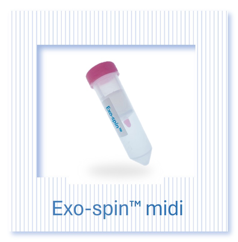
Exo-spin SEC isolation kits. For small to medium conditioned media volumes, Exo-spin size exclusion chromatography kits provide high-purity EV isolation that separates vesicles from co-isolated proteins by size. Available in mini, midi and maxi formats for volumes from 100 microlitres to 50 ml, with a validated protocol suitable for cell conditioned media from all common cell types.
EVlution TFF system. For large conditioned media volumes from bioreactor or multi-layer flask cultures, the EVlution tangential flow filtration system concentrates EV preparations from several litres of conditioned media in a single continuous run. Switch Flow technology enables retentate recovery below 10 ml from the closed-path, disposable sample-contact circuit.
ExoLISA quantitative tetraspanin assays. ExoLISA CD9, CD63 and CD81 assays provide plate-based quantitative tetraspanin detection for MISEV2018-compliant characterisation without the inter-operator variability of western blot. Suitable for routine inter-lot comparison of EV preparations.
NTA size profiling service. The Cell Guidance Systems NTA size profiling service provides MISEV-compliant particle concentration and size distribution analysis for submitted EV preparations, supporting laboratories without in-house NTA instrumentation.
EV and exosome characterisation services. Full MISEV2018-compliant characterisation including NTA, tetraspanin profiling and electron microscopy is available as part of the Cell Guidance Systems EV and Exosome Services package.
| EV Research Starting Point: Decision Guide | |
| Starting point | Recommended approach |
| Conditioned media under 50 ml, research-scale isolation | Exo-spin SEC isolation |
| Large conditioned media volume or bioreactor output | EVlution TFF followed by Exo-spin SEC |
| MISEV tetraspanin quantification across preparation lots | ExoLISA assays (CD9, CD63, CD81) |
| No in-house NTA capability | CGS NTA size profiling service |
| Full MISEV characterisation package required | CGS EV and Exosome Services |
Understanding what extracellular vesicles are, and which type you are actually working with, is the foundation on which all downstream EV research depends. The next article in this series examines how the major isolation methods, ultracentrifugation, size exclusion chromatography and tangential flow filtration, compare in terms of purity, yield and practical applicability across different experimental settings. For researchers already working with isolated EV preparations, our article on cancer diagnostic EVs and our detailed review of MSC-derived exosomes in regenerative medicine provide applied context for the biology introduced here.
References
[1] Pan BT, Johnstone RM. Fate of the transferrin receptor during maturation of sheep reticulocytes in vitro: selective externalization of the receptor. Cell. 1983;33(3):967-978.
[2] Johnstone RM, Adam M, Hammond JR, Orr L, Turbide C. Vesicle formation during reticulocyte maturation. Association of plasma membrane activities with released vesicles (exosomes). J Biol Chem. 1987;262(19):9412-9420.
[3] Raposo G, Stoorvogel W. Extracellular vesicles: exosomes, microvesicles, and friends. J Cell Biol. 2013;200(4):373-383.
[4] van Niel G, D'Angelo G, Raposo G. Shedding light on the cell biology of extracellular vesicles. Nat Rev Mol Cell Biol. 2018;19(4):213-228.
[5] MISEV2023: a position statement of the International Society for Extracellular Vesicles and update of the MISEV2014 and MISEV 2018 guidelines.
[6] Kalluri R, LeBleu VS. The biology, function, and biomedical applications of exosomes. Science. 2020;367(6478):eaau6977.
[7] Mathieu M, Martin-Jaular L, Lavieu G, Théry C. Specificities of secretion and uptake of exosomes and other extracellular vesicles for cell-to-cell communication. Nat Cell Biol. 2019;21(1):9-17.