Exosomes vs liposomes for drug delivery.

Exosomes and liposomes viewed under an electron microscope may look very similar: Both are spherical, lipid bilayer-enclosed particles in the tens-to-hundreds of nanometres range, both carry an aqueous interior, and both can deliver a molecular payload into a cell. Yet as drug delivery platforms, they occupy very different positions. Liposomes are a fifty-year-old, clinically approved, industrially manufactured technology. Exosomes are a natural product of cell biology that the field is still learning to produce, load and characterise reliably.
Choosing between them is not a matter of deciding which is intrinsically better. It is a matter of understanding what each platform does well and badly, and which of those strengths and weaknesses matter for a particular therapeutic problem. This article compares the two across the criteria that actually determine project outcomes: cargo loading, targeting, immune handling, manufacturability, analytical control and regulatory precedent.
Origins
The single most important difference between exosomes and liposomes is not structural. It is that one is designed and the other is biomimetic
A liposome is assembled from a defined list of components. A formulation scientist selects phospholipids, typically a phosphatidylcholine such as hydrogenated soy PC, adds cholesterol to modulate membrane rigidity, and often incorporates a PEGylated lipid to extend circulation time. Composition is a specification. Every molecule present is there because someone chose to put it there, and the same formulation can be reproduced indefinitely.
An exosome is not assembled, it is secreted. Exosomes are a subtype of extracellular vesicle (EV) formed by inward budding of the endosomal membrane to create intraluminal vesicles within multivesicular bodies, which are then released when the multivesicular body fuses with the plasma membrane. Their composition is set by the biology of the producing cell: the lipid profile is enriched in cholesterol, sphingomyelin and phosphatidylserine relative to the plasma membrane, the surface carries tetraspanins such as CD9, CD63 and CD81 alongside integrins, adhesion molecules and a protein corona, and the lumen contains a sample of the parent cell's RNA and protein content. None of this is specified. It is measured after the fact.
That distinction propagates into every downstream decision. Liposomes are engineered from a blank sheet and must have every desirable property added deliberately. Exosomes arrive with a large set of biological properties already present, most of which are useful, some of which are not, and few of which can be turned off.
A Note on Liposomes vs Lipid Nanoparticles
Liposomes and lipid nanoparticles (LNPs) are frequently conflated, and the distinction matters for any comparison with exosomes. A classical liposome has a lamellar structure: one or more concentric bilayers surrounding an aqueous core in which hydrophilic cargo is dissolved. An LNP, the format used in patisiran and in the mRNA vaccines, is not lamellar. It has an electron-dense core in which ionisable cationic lipids complex nucleic acid cargo directly, with no discrete aqueous compartment.
The functional consequence is significant. LNPs deliver nucleic acids efficiently because the ionisable lipid becomes protonated in the acidifying endosome and destabilises the endosomal membrane, driving cargo release into the cytosol. Classical liposomes have no such mechanism and are generally poor at cytosolic delivery of nucleic acids. When the literature compares exosomes favourably with "liposomes" for RNA delivery, the comparator is often a classical liposome rather than a modern LNP, and the comparison should be read with that in mind.
Side-by-Side: An Interactive Comparison
The panel below compares the two platforms across nine criteria. The scores are qualitative indicators of relative standing based on the published literature and current manufacturing practice, not measured values. Select a criterion to see how the platforms compare and why.
Figure: Structural comparison and relative platform scoring. Scores are qualitative and illustrative, intended to summarise consensus positions in the literature rather than to report measured values.
Cargo Loading: The Clearest Liposome Advantage
Hydrophilic drugs can be dissolved in the aqueous phase during hydration and become encapsulated as vesicles form. Lipophilic drugs partition into the bilayer. Most powerfully, amphipathic weak bases such as doxorubicin can be remotely loaded across a preformed transmembrane gradient: the drug diffuses in as a neutral species, becomes protonated in the acidic interior, and precipitates as a gel that cannot diffuse back out. Encapsulation efficiencies above 90% are routine, and drug-to-lipid ratios can be specified rather than measured.
Exosome loading has no equivalent. The vesicle already exists when the cargo is introduced, and its membrane is not designed to be permeabilised. The available options fall into two groups. Exogenous methods, electroporation, sonication, saponin treatment, freeze-thaw cycling and chemical transfection, force cargo across an intact membrane and generally achieve low single-digit to low double-digit percentage loading, often with vesicle aggregation or membrane damage as a side effect. Endogenous methods engineer the producer cell to overexpress the cargo or to fuse it to an EV-sorting protein, which avoids post-isolation stress but constrains what can be loaded to what a cell can be persuaded to make and package.
For small-molecule drugs, this gap is decisive and liposomes win based on loading alone. For nucleic acids and proteins where the cargo can be expressed by the producer cell, endogenous exosome loading becomes competitive, and the argument shifts more to what happens after the vesicle reaches the target cell.
Targeting and Biodistribution
An untargeted liposome injected intravenously accumulates predominantly in liver and spleen through mononuclear phagocyte system uptake. PEGylation reduces opsonisation and extends circulation half-life from minutes to many hours, which allows passive accumulation in tissues with leaky vasculature. Active targeting, achieved by conjugating antibodies, antibody fragments, peptides or aptamers to the liposome surface, has produced impressive preclinical results but has translated to the clinic only sporadically. The practical difficulty is that adding a targeting ligand also adds a recognition surface for the immune system and complicates manufacturing and release testing.
Exosomes start from a different position. Their surfaces carry integrins, tetraspanins, adhesion molecules and glycans acquired from the producer cell, and there is evidence that this composition influences where they go. Hoshino and colleagues reported that tumour-derived exosome integrin profiles correlated with organotropic metastatic patterns, with specific integrin combinations associating with lung, liver or brain destinations. Wiklander and colleagues showed that EV biodistribution in mice depends on cell source, administration route and targeting modification, indicating that producer cell choice is itself a design lever.
Exosome tropism is real but probably modest, and the majority of an intravenously administered dose still ends up in liver and spleen. Exosomes offer a biologically informed starting point rather than solving the targeting problem. Where deliberate, efficient targeting is required, engineered display remains the approach: the classic demonstration by Alvarez-Erviti and colleagues fused the rabies virus glycoprotein peptide to Lamp2b to direct siRNA-loaded exosomes to mouse brain after systemic injection.
Immune Handling and Repeat Dosing
PEGylation is the standard liposome answer to rapid clearance, and it works, but it carries a documented complication. Repeat administration of PEGylated liposomes can induce anti-PEG antibodies and the accelerated blood clearance phenomenon, in which a second dose is cleared substantially faster than the first. Complement activation-related pseudoallergy is a further recognised infusion reaction associated with some liposomal products.
Exosomes carry endogenous immunomodulatory surface features, most notably CD47, which engages SIRP-alpha on phagocytes and acts as a signal to limit phagocytosis. Kamerkar and colleagues attributed the superior circulation retention of exosomes over liposomes in their pancreatic cancer model to exactly this mechanism, and reported CD47-dependent efficacy for siRNA-loaded exosomes targeting mutant KRAS.
The counterbalancing risk is that a biologically derived particle carries biologically active material that was not deliberately included. Producer cell proteins, residual medium components, co-purified lipoproteins and, for some sources, procoagulant phosphatidylserine exposure all require assessment. Liposome immunotoxicology is comparatively well mapped. Exosome immunotoxicology is source-specific and must be established for each production system.
Head-to-Head Comparison
| Exosomes and liposomes compared across key delivery criteria | ||
| Criterion | Liposomes | Exosomes |
| Origin | Synthetic, assembled from defined lipids | Biological, secreted via the endosomal pathway |
| Typical size | 50 to 200 nm, tunable by extrusion | 30 to 200 nm, polydisperse and source-dependent |
| Composition control | Complete, set by formulation | Indirect, via producer cell and culture conditions |
| Cargo loading | Passive and remote loading, often above 90% efficiency | Electroporation, sonication or producer cell engineering, typically low and variable |
| Targeting | Passive accumulation, or added ligands | Native tropism from surface integrins and tetraspanins, extendable by display engineering |
| Circulation strategy | PEG steric stabilisation, with anti-PEG and ABC risk | Endogenous CD47 and other self markers |
| Manufacturing | Microfluidic mixing or extrusion, scales predictably | Cell culture plus TFF, SEC and polishing, yield-limited |
| Storage | Established liquid and lyophilised formulations | Commonly minus 80 degrees C, with lyophilisation under active development |
| Regulatory status | Multiple approved products since 1995 | No approved therapeutic to date, numerous clinical trials ongoing |
Manufacturing and Analytical Control
This is where the practical gap between the platforms is widest, and where most exosome programmes encounter their real difficulties.
A liposome batch is defined by its recipe. Analytical release involves lipid identity and content, particle size and polydispersity, zeta potential, encapsulated and free drug, and sterility. Every one of these is a mature assay with clear acceptance criteria.
An exosome batch is defined by what came out of a bioreactor. Its identity has to be demonstrated rather than specified, which under MISEV2023 means quantifying particle number, demonstrating the presence of transmembrane and cytosolic EV markers, showing the absence or acceptable level of non-EV contaminants, and reporting the isolation method in sufficient detail for others to reproduce it. Particle-to-protein ratio, tetraspanin marker profiles by immunoassay, and size distribution by nanoparticle tracking analysis all become routine release requirements rather than optional characterisation.
Purification methodology matters correspondingly more. Differential ultracentrifugation, still widely used in research, is poorly suited to scale and causes vesicle damage and aggregation. Tangential flow filtration (TFF) combined with size exclusion chromatography has become the preferred route for larger preparations because it concentrates gently, permits buffer exchange by diafiltration, and scales with membrane area. Getting this process right is not a peripheral concern: for an EV therapeutic, the purification process is a substantial part of the product definition.
Hybrid Approaches
Given that the two platforms have close to complementary strengths, hybrid vesicles are an obvious idea and an active area of work. Fusing exosome membranes with liposomes, typically by freeze-thaw cycling or by incubation with membrane-destabilising agents, produces particles that retain exosomal surface proteins while allowing synthetic lipids and established loading chemistries to be introduced. The stated goal is exosomal targeting and immune handling with liposomal loading efficiency and manufacturability.
Hybrids are promising but should be assessed carefully. Characterising a fused particle population is harder than characterising either parent, incomplete fusion produces mixed populations that are difficult to resolve analytically, and the regulatory position for a part-biological, part-synthetic nanoparticle is undefined. The concept is sound. The analytical and manufacturing burden is higher than either single platform, not lower.
Choosing Between Them
| Which platform fits which problem | |
| Situation | Preferred starting point |
| Small-molecule drug requiring high loading and defined dose per particle | Liposome, where remote loading gives control that exosomes cannot match |
| Nucleic acid requiring cytosolic delivery at scale | Lipid nanoparticle, with exosomes considered where LNP hepatotropism or reactogenicity is limiting |
| Therapeutic effect derived from the vesicle itself rather than an added payload | Exosome, since the biological cargo is the product |
| Repeat chronic dosing where anti-PEG responses are a concern | Exosome, or a non-PEGylated liposome formulation |
| Delivery to tissue that is poorly accessed by conventional nanoparticles | Exosome, exploiting producer cell selection and surface display engineering |
| Programme constrained by cost of goods or near-term regulatory timeline | Liposome, where precedent and manufacturing maturity substantially de-risk development |
Tools for EV Delivery Research
Any exosome delivery programme depends on producing well-characterised vesicles reproducibly, which in practice means getting purification and analytics right before making claims about delivery. Cell Guidance Systems supplies tools across that workflow.
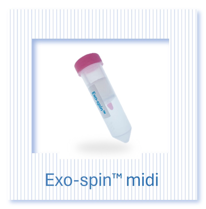
The EVlution TFF system supports gentle concentration and diafiltration of EV preparations using hollow fibre cartridges, the approach best suited to scaling beyond what ultracentrifugation can handle without damaging vesicles. For smaller preparations, Exo-spin columns combine precipitation and size exclusion chromatography in a research-scale format.
On the characterisation side, ExoLISA assays provide quantitative detection of EV surface markers including the tetraspanins expected under MISEV2023 identity criteria, and NTA size profiling is available as a service for particle concentration and size distribution measurement. Groups without in-house instrumentation can access processing and analysis through our EV and exosome services, and custom freeze dried EVs are available for groups investigating alternatives to frozen storage.
Summary
Liposomes and exosomes are not competing answers to the same question. Liposomes are a mature pharmaceutical technology with unmatched control over composition, loading and manufacture, and with three decades of regulatory precedent behind them. Exosomes are a biological delivery system that arrives pre-equipped with tropism, immune compatibility and cytosolic delivery mechanisms that liposome formulators have spent decades trying to reproduce synthetically, but which is still difficult to load, expensive to produce and demanding to characterise.
The sensible reading is that liposomes remain the default for defined small-molecule payloads and for programmes where manufacturing and regulatory risk must be minimised, that lipid nanoparticles remain the default for nucleic acid delivery at scale, and that exosomes are the right choice where the biology of the vesicle is doing part of the therapeutic work. As EV manufacturing and analytics mature, the boundary will move. It has not moved yet, and being clear-eyed about where it currently sits is the most useful thing a delivery programme can do at the outset.
Further Reading
- Welsh JA, et al. Minimal information for studies of extracellular vesicles (MISEV2023): from basic to advanced approaches. J Extracell Vesicles. 2024;13(2):e12404.
- van der Meel R, et al. Extracellular vesicles as drug delivery systems: lessons from the liposome field. J Control Release. 2014;195:72-85.
- Murphy DE, et al. Extracellular vesicle-based therapeutics: natural versus engineered targeting and trafficking. Exp Mol Med. 2019;51(3):1-12.
- Kamerkar S, et al. Exosomes facilitate therapeutic targeting of oncogenic KRAS in pancreatic cancer. Nature. 2017;546(7659):498-503.
- Alvarez-Erviti L, et al. Delivery of siRNA to the mouse brain by systemic injection of targeted exosomes. Nat Biotechnol. 2011;29(4):341-345.
- Hoshino A, et al. Tumour exosome integrins determine organotropic metastasis. Nature. 2015;527(7578):329-335.
- Wiklander OPB, et al. Extracellular vesicle in vivo biodistribution is determined by cell source, route of administration and targeting. J Extracell Vesicles. 2015;4:26316.
- Sercombe L, et al. Advances and challenges of liposome assisted drug delivery. Front Pharmacol. 2015;6:286.
- Barenholz Y. Doxil, the first FDA-approved nano-drug: lessons learned. J Control Release. 2012;160(2):117-134.
- Adams D, et al. Patisiran, an RNAi therapeutic, for hereditary transthyretin amyloidosis. N Engl J Med. 2018;379(1):11-21.
Learn more about EV purification and characterisation tools from Cell Guidance Systems: